 Movie
Movie Controller

[English] 日本語
 Yorodumi
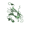
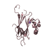
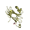
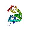
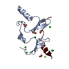
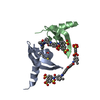
- PDB-3qec: Crystal structure of a putative carbohydrate binding protein (PA1... -
+ Open data

- Basic information
| Entry | Database: PDB / ID: 3qec | ||||||
|---|---|---|---|---|---|---|---|
| Title | Crystal structure of a putative carbohydrate binding protein (PA1324) from Pseudomonas aeruginosa at 2.61 A resolution | ||||||
 Components Components | Putative carbohydrate binding protein | ||||||
 Keywords Keywords | CARBOHYDRATE-BINDING PROTEIN / SURAMIN BINDING / HEPARIN BINDING / POSSIBLE CARBOHYDRATE TRANSPORTER / BIOFILM FORMATION / STRUCTURAL GENOMICS / JOINT CENTER FOR STRUCTURAL GENOMICS / JCSG / PROTEIN STRUCTURE INITIATIVE / PSI-BIOLOGY / CARBOHYDRATE- BINDING PROTEIN | ||||||
| Function / homology | Prokaryotic membrane lipoprotein lipid attachment site profile. / DI(HYDROXYETHYL)ETHER / TRIETHYLENE GLYCOL / Carboxypeptidase regulatory-like domain-containing protein Function and homology information Function and homology information | ||||||
| Biological species |   Pseudomonas aeruginosa (bacteria) Pseudomonas aeruginosa (bacteria) | ||||||
| Method |  X-RAY DIFFRACTION / SYNCHROTRON / MAD / Resolution: 2.61 Å X-RAY DIFFRACTION / SYNCHROTRON / MAD / Resolution: 2.61 Å | ||||||
 Authors Authors | Joint Center for Structural Genomics (JCSG) | ||||||
 Citation Citation | Journal: To be published Title: Crystal structure of a putative carbohydrate binding protein (PA1324) from Pseudomonas aeruginosa at 2.61 A resolution Authors: Joint Center for Structural Genomics (JCSG) | ||||||
| History |
|
- Structure visualization
| Structure viewer | Molecule: MolmilJmol/JSmol |
|---|
- Downloads & links
-Download
| PDBx/mmCIF format | 3qec.cif.gz | 73.7 KB | Display | PDBx/mmCIF format |
|---|---|---|---|---|
| PDB format | pdb3qec.ent.gz | 55.1 KB | Display | PDB format |
| PDBx/mmJSON format | 3qec.json.gz | Tree view | PDBx/mmJSON format | |
| Others |  Other downloads Other downloads |
-Validation report
| Arichive directory | https://data.pdbj.org/pub/pdb/validation_reports/qe/3qecftp://data.pdbj.org/pub/pdb/validation_reports/qe/3qec | HTTPS FTP |
|---|
-Related structure data
| Similar structure data | |
|---|---|
| Other databases |


-Links
 PDBj
PDBj


- Assembly
| Deposited unit | 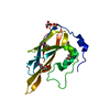
| |||||||||
|---|---|---|---|---|---|---|---|---|---|---|
| 1 |
| |||||||||
| 2 | 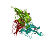
| |||||||||
| Unit cell |
| |||||||||
| Components on special symmetry positions |
| |||||||||
| Details | CRYSTAL PACKING ANALYSIS SUPPORTS THE ASSIGNMENT OF A MONOMER AS THE SIGNIFICANT OLIGOMERIC FORM. |
-Components
-Protein , 1 types, 1 molecules A
| #1: Protein | Mass: 16336.757 Da / Num. of mol.: 1 / Fragment: sequence database residues 22-170 Source method: isolated from a genetically manipulated source Source: (gene. exp.) Pseudomonas aeruginosa (bacteria) / Gene: PA1324 / Plasmid: SpeedET / Production host: |
|---|
-Non-polymers , 5 types, 45 molecules 


| #2: Chemical | ChemComp-SO4 /  Mass: 96.063 Da / Num. of mol.: 1 / Source method: obtained synthetically / Formula: SO4 Mass: 96.063 Da / Num. of mol.: 1 / Source method: obtained synthetically / Formula: SO4 | ||||||
|---|---|---|---|---|---|---|---|
| #3: Chemical |  Mass: 92.094 Da / Num. of mol.: 2 / Source method: obtained synthetically / Formula: C3H8O3 Mass: 92.094 Da / Num. of mol.: 2 / Source method: obtained synthetically / Formula: C3H8O3#4: Chemical |  Mass: 106.120 Da / Num. of mol.: 2 / Source method: obtained synthetically / Formula: C4H10O3 Mass: 106.120 Da / Num. of mol.: 2 / Source method: obtained synthetically / Formula: C4H10O3#5: Chemical | ChemComp-PGE / |  Mass: 150.173 Da / Num. of mol.: 1 / Source method: obtained synthetically / Formula: C6H14O4 Mass: 150.173 Da / Num. of mol.: 1 / Source method: obtained synthetically / Formula: C6H14O4#6: Water | ChemComp-HOH / | Mass: 18.015 Da / Num. of mol.: 39 / Source method: isolated from a natural source / Formula: H2O |
-Details
| Has protein modification | Y |
|---|---|
| Sequence details | THE CONSTRUCT (RESIDUES 22-170) WAS EXPRESSED WITH A PURIFICATION TAG MGSDKIHHHHHHENLYFQG. THE TAG ...THE CONSTRUCT (RESIDUES 22-170) WAS EXPRESSED WITH A PURIFICATI |
-Experimental details
-Experiment
| Experiment | Method: X-RAY DIFFRACTION / Number of used crystals: 1 |
|---|
- Sample preparation
| Crystal grow | Temperature: 293 K / Method: vapor diffusion, sitting drop / pH: 7.9 Details: 15.00% Glycerol, 1.85% polyethylene glycol 400, 2.10M ammonium sulfate, 0.1M HEPES pH 7.9, NANODROP', VAPOR DIFFUSION, SITTING DROP, temperature 293K |
|---|
-Data collection
| Diffraction | Mean temperature: 100 K | |||||||||||||||||||||||||||||||||||||||||||||||||||||||||||||||||||||||||||||
|---|---|---|---|---|---|---|---|---|---|---|---|---|---|---|---|---|---|---|---|---|---|---|---|---|---|---|---|---|---|---|---|---|---|---|---|---|---|---|---|---|---|---|---|---|---|---|---|---|---|---|---|---|---|---|---|---|---|---|---|---|---|---|---|---|---|---|---|---|---|---|---|---|---|---|---|---|---|---|
| Diffraction source | Source: SYNCHROTRON / Site: SSRL  / Beamline: BL11-1 / Wavelength: 0.91837,0.98012,0.97944 / Beamline: BL11-1 / Wavelength: 0.91837,0.98012,0.97944 | |||||||||||||||||||||||||||||||||||||||||||||||||||||||||||||||||||||||||||||
| Detector | Type: MARMOSAIC 325 mm CCD / Detector: CCD / Date: Nov 23, 2010 | |||||||||||||||||||||||||||||||||||||||||||||||||||||||||||||||||||||||||||||
| Radiation | Protocol: MAD / Monochromatic (M) / Laue (L): M / Scattering type: x-ray | |||||||||||||||||||||||||||||||||||||||||||||||||||||||||||||||||||||||||||||
| Radiation wavelength |
| |||||||||||||||||||||||||||||||||||||||||||||||||||||||||||||||||||||||||||||
| Reflection | Resolution: 2.61→29.692 Å / Num. obs: 12763 / % possible obs: 97.6 % / Observed criterion σ(I): -3 / Biso Wilson estimate: 84.108 Å2 / Rmerge(I) obs: 0.054 / Net I/σ(I): 28.75 | |||||||||||||||||||||||||||||||||||||||||||||||||||||||||||||||||||||||||||||
| Reflection shell |
|
-Phasing
| Phasing | Method: MAD |
|---|
- Processing
| Software |
| ||||||||||||||||||||||||||||||||||||||||||||||||||||||||||||||||||||||||||||||||||||||||||||||||||||||||||||
|---|---|---|---|---|---|---|---|---|---|---|---|---|---|---|---|---|---|---|---|---|---|---|---|---|---|---|---|---|---|---|---|---|---|---|---|---|---|---|---|---|---|---|---|---|---|---|---|---|---|---|---|---|---|---|---|---|---|---|---|---|---|---|---|---|---|---|---|---|---|---|---|---|---|---|---|---|---|---|---|---|---|---|---|---|---|---|---|---|---|---|---|---|---|---|---|---|---|---|---|---|---|---|---|---|---|---|---|---|---|
| Refinement | Method to determine structure: MAD / Resolution: 2.61→29.692 Å / Cor.coef. Fo:Fc: 0.9345 / Cor.coef. Fo:Fc free: 0.9339 / Occupancy max: 1 / Occupancy min: 0.33 / Cross valid method: THROUGHOUT / σ(F): 0 Details: 1. A MET-INHIBITION PROTOCOL WAS USED FOR SELENOMETHIONINE INCORPORATION DURING PROTEIN EXPRESSION. THE OCCUPANCY OF THE SE ATOMS IN THE MSE RESIDUES WAS REDUCED TO 0.75 FOR THE REDUCED ...Details: 1. A MET-INHIBITION PROTOCOL WAS USED FOR SELENOMETHIONINE INCORPORATION DURING PROTEIN EXPRESSION. THE OCCUPANCY OF THE SE ATOMS IN THE MSE RESIDUES WAS REDUCED TO 0.75 FOR THE REDUCED SCATTERING POWER DUE TO PARTIAL S-MET INCORPORATION. 2. ATOM RECORD CONTAINS SUM OF TLS AND RESIDUAL B FACTORS. ANISOU RECORD CONTAINS SUM OF TLS AND RESIDUAL U FACTORS. 3. SULFATE (SO4) AND PEG-400 FRAGMENTS (PEG AND PGE) FROM THE CRYSTALLIZATION SOLUTION AND GLYCEROL FROM THE CRYOPROTECTANT HAVE BEEN MODELED IN THE SOLVENT STRUCTURE.
| ||||||||||||||||||||||||||||||||||||||||||||||||||||||||||||||||||||||||||||||||||||||||||||||||||||||||||||
| Displacement parameters | Biso max: 156.18 Å2 / Biso mean: 70.3766 Å2 / Biso min: 45.68 Å2
| ||||||||||||||||||||||||||||||||||||||||||||||||||||||||||||||||||||||||||||||||||||||||||||||||||||||||||||
| Refinement step | Cycle: LAST / Resolution: 2.61→29.692 Å
| ||||||||||||||||||||||||||||||||||||||||||||||||||||||||||||||||||||||||||||||||||||||||||||||||||||||||||||
| Refine LS restraints |
| ||||||||||||||||||||||||||||||||||||||||||||||||||||||||||||||||||||||||||||||||||||||||||||||||||||||||||||
| LS refinement shell | Resolution: 2.61→2.86 Å / Total num. of bins used: 6
| ||||||||||||||||||||||||||||||||||||||||||||||||||||||||||||||||||||||||||||||||||||||||||||||||||||||||||||
| Refinement TLS params. | Method: refined / Origin x: 108.969 Å / Origin y: 133.414 Å / Origin z: 125.085 Å
|
