 Movie
Movie Controller

[English] 日本語
 Yorodumi
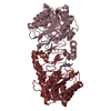
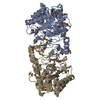
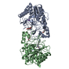
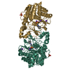
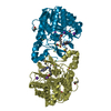
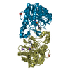
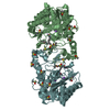
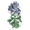
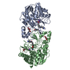
- PDB-3q9c: Crystal Structure of H159A APAH complexed with N8-acetylspermidine -
+ Open data

- Basic information
| Entry | Database: PDB / ID: 3q9c | ||||||
|---|---|---|---|---|---|---|---|
| Title | Crystal Structure of H159A APAH complexed with N8-acetylspermidine | ||||||
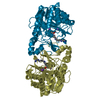
 Components Components | Acetylpolyamine amidohydrolase | ||||||
 Keywords Keywords | HYDROLASE / HDAC / polyamine / Arginase Fold / Deacetylase | ||||||
| Function / homology |  Function and homology information Function and homology informationacetylspermidine deacetylase / acetylspermidine deacetylase activity / acetylputrescine deacetylase / acetylputrescine deacetylase activity / histone deacetylase activity / Hydrolases; Acting on carbon-nitrogen bonds, other than peptide bonds; In linear amides / epigenetic regulation of gene expression / metal ion binding Similarity search - Function | ||||||
| Biological species |  Mycoplana ramosa (bacteria) Mycoplana ramosa (bacteria) | ||||||
| Method |  X-RAY DIFFRACTION / SYNCHROTRON / FOURIER SYNTHESIS / Resolution: 2.3 Å X-RAY DIFFRACTION / SYNCHROTRON / FOURIER SYNTHESIS / Resolution: 2.3 Å | ||||||
 Authors Authors | Lombardi, P.M. / Christianson, D.W. | ||||||
 Citation Citation | Journal: Biochemistry / Year: 2011 Title: Structure of prokaryotic polyamine deacetylase reveals evolutionary functional relationships with eukaryotic histone deacetylases . Authors: Lombardi, P.M. / Angell, H.D. / Whittington, D.A. / Flynn, E.F. / Rajashankar, K.R. / Christianson, D.W. | ||||||
| History |
|
- Structure visualization
| Structure viewer | Molecule: MolmilJmol/JSmol |
|---|
- Downloads & links
-Download
| PDBx/mmCIF format | 3q9c.cif.gz | 785.1 KB | Display | PDBx/mmCIF format |
|---|---|---|---|---|
| PDB format | pdb3q9c.ent.gz | 649 KB | Display | PDB format |
| PDBx/mmJSON format | 3q9c.json.gz | Tree view | PDBx/mmJSON format | |
| Others |  Other downloads Other downloads |
-Validation report
| Arichive directory | https://data.pdbj.org/pub/pdb/validation_reports/q9/3q9cftp://data.pdbj.org/pub/pdb/validation_reports/q9/3q9c | HTTPS FTP |
|---|
-Related structure data
| Related structure data |  3q9bC  3q9eC 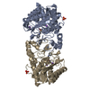 3q9fSC C: citing same article ( S: Starting model for refinement |
|---|---|
| Similar structure data |


-Links
 PDBj
PDBj- Assembly
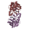
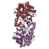
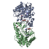
| Deposited unit | 
| ||||||||
|---|---|---|---|---|---|---|---|---|---|
| 1 | 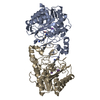
| ||||||||
| 2 | 
| ||||||||
| 3 | 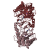
| ||||||||
| 4 | 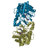
| ||||||||
| 5 | 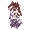
| ||||||||
| 6 | 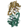
| ||||||||
| Unit cell |
|
-Components
-Protein , 1 types, 12 molecules ABCDEFGHIJKL
| #1: Protein | Mass: 36302.926 Da / Num. of mol.: 12 / Mutation: H159A Source method: isolated from a genetically manipulated source Source: (gene. exp.) Mycoplana ramosa (bacteria) / Gene: aphA, aph / Plasmid: pET-21b / Production host: |
|---|
-Non-polymers , 5 types, 1800 molecules 


| #2: Chemical | ChemComp-Q9C /  Mass: 187.283 Da / Num. of mol.: 12 / Source method: obtained synthetically / Formula: C9H21N3O Mass: 187.283 Da / Num. of mol.: 12 / Source method: obtained synthetically / Formula: C9H21N3O#3: Chemical | ChemComp-K /  Mass: 39.098 Da / Num. of mol.: 12 / Source method: obtained synthetically / Formula: K Mass: 39.098 Da / Num. of mol.: 12 / Source method: obtained synthetically / Formula: K#4: Chemical | ChemComp-ZN /  Mass: 65.409 Da / Num. of mol.: 12 / Source method: obtained synthetically / Formula: Zn Mass: 65.409 Da / Num. of mol.: 12 / Source method: obtained synthetically / Formula: Zn#5: Chemical | ChemComp-NA /  Mass: 22.990 Da / Num. of mol.: 12 / Source method: obtained synthetically / Formula: Na Mass: 22.990 Da / Num. of mol.: 12 / Source method: obtained synthetically / Formula: Na#6: Water | ChemComp-HOH / | Mass: 18.015 Da / Num. of mol.: 1752 / Source method: isolated from a natural source / Formula: H2O |
|---|
-Experimental details
-Experiment
| Experiment | Method: X-RAY DIFFRACTION / Number of used crystals: 1 |
|---|
- Sample preparation
| Crystal | Density Matthews: 3.37 Å3/Da / Density % sol: 63.52 % |
|---|---|
| Crystal grow | Temperature: 298 K / Method: vapor diffusion, hanging drop / pH: 6.2 Details: 1.0 M sodium phosphate, 0.8 M potassium phosphate, 0.2 M lithium sulfate, 0.1 M CAPS pH 6.2, VAPOR DIFFUSION, HANGING DROP, temperature 298K |
-Data collection
| Diffraction | Mean temperature: 100 K |
|---|---|
| Diffraction source | Source: SYNCHROTRON / Site: APS  / Beamline: 24-ID-C / Wavelength: 0.97949 Å / Beamline: 24-ID-C / Wavelength: 0.97949 Å |
| Detector | Type: ADSC QUANTUM 315r / Detector: CCD / Date: Jun 11, 2010 / Details: Kirpatrick Baez focusing mirrors |
| Radiation | Monochromator: Si(111) double crystal monochromator / Protocol: SINGLE WAVELENGTH / Monochromatic (M) / Laue (L): M / Scattering type: x-ray |
| Radiation wavelength | Wavelength: 0.97949 Å / Relative weight: 1 |
| Reflection | Resolution: 2.3→50 Å / Num. all: 252823 / Num. obs: 219596 / % possible obs: 86.9 % / Observed criterion σ(F): 0 / Observed criterion σ(I): -3 / Redundancy: 2.4 % / Rmerge(I) obs: 0.109 / Rsym value: 0.109 / Net I/σ(I): 7.472 |
| Reflection shell | Resolution: 2.3→2.37 Å / Redundancy: 2.3 % / Rmerge(I) obs: 0.361 / Mean I/σ(I) obs: 2.059 / Num. unique all: 21913 / Rsym value: 0.361 / % possible all: 96.9 |
- Processing
| Software |
| ||||||||||||||||||||||||||||||||
|---|---|---|---|---|---|---|---|---|---|---|---|---|---|---|---|---|---|---|---|---|---|---|---|---|---|---|---|---|---|---|---|---|---|
| Refinement | Method to determine structure: FOURIER SYNTHESIS Starting model: PDB ENTRY 3Q9F Resolution: 2.3→50 Å / Occupancy max: 1 / Occupancy min: 1 / Cross valid method: THROUGHOUT / σ(F): 0 / Stereochemistry target values: Engh & Huber
| ||||||||||||||||||||||||||||||||
| Solvent computation | Bsol: 47.0561 Å2 | ||||||||||||||||||||||||||||||||
| Displacement parameters | Biso max: 105.52 Å2 / Biso mean: 25.677 Å2 / Biso min: 4.33 Å2
| ||||||||||||||||||||||||||||||||
| Refine analyze |
| ||||||||||||||||||||||||||||||||
| Refinement step | Cycle: LAST / Resolution: 2.3→50 Å
| ||||||||||||||||||||||||||||||||
| Refine LS restraints |
| ||||||||||||||||||||||||||||||||
| LS refinement shell | Resolution: 2.3→2.38 Å
| ||||||||||||||||||||||||||||||||
| Xplor file |
|
