 Movie
Movie Controller


+ Open data

- Basic information
| Entry | Database: PDB / ID: 3q5c | ||||||||||||||||||
|---|---|---|---|---|---|---|---|---|---|---|---|---|---|---|---|---|---|---|---|
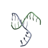
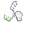
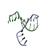
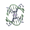
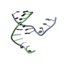
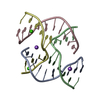
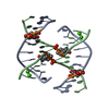
| Title | Crystal structure of four-way junction with sticky end | ||||||||||||||||||
 Components Components | DNA (5'-D(* Keywords KeywordsDNA / Sticky end | Function / homology | DNA |  Function and homology information Function and homology informationMethod |  X-RAY DIFFRACTION / SYNCHROTRON / MOLECULAR REPLACEMENT / Resolution: 2.501 Å X-RAY DIFFRACTION / SYNCHROTRON / MOLECULAR REPLACEMENT / Resolution: 2.501 Å  Authors AuthorsVenkadesh, S. / Mandal, P.K. / Gautham, N. |  CitationJournal: Biochem.Biophys.Res.Commun. / Year: 2011 CitationJournal: Biochem.Biophys.Res.Commun. / Year: 2011Title: The sequence d(CGGCGGCCGC) self-assembles into a two dimensional rhombic DNA lattice Authors: Venkadesh, S. / Mandal, P.K. / Gautham, N. History |
|
- Structure visualization
| Structure viewer | Molecule: MolmilJmol/JSmol |
|---|
- Downloads & links
-Download
| PDBx/mmCIF format | 3q5c.cif.gz | 29.6 KB | Display | PDBx/mmCIF format |
|---|---|---|---|---|
| PDB format | pdb3q5c.ent.gz | 20.5 KB | Display | PDB format |
| PDBx/mmJSON format | 3q5c.json.gz | Tree view | PDBx/mmJSON format | |
| Others |  Other downloads Other downloads |
-Validation report
| Arichive directory | https://data.pdbj.org/pub/pdb/validation_reports/q5/3q5cftp://data.pdbj.org/pub/pdb/validation_reports/q5/3q5c | HTTPS FTP |
|---|
-Related structure data
| Related structure data |  1p4yS S: Starting model for refinement |
|---|---|
| Similar structure data |

-Links
 PDBj
PDBj

- Assembly
| Deposited unit | 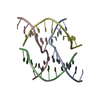
| ||||||||
|---|---|---|---|---|---|---|---|---|---|
| 1 |
| ||||||||
| Unit cell |
|
-Components
| #1: DNA chain | Mass: 3046.980 Da / Num. of mol.: 4 / Source method: obtained synthetically / Details: Chemically synthesized #2: Water | ChemComp-HOH / |  Mass: 18.015 Da / Num. of mol.: 9 / Source method: isolated from a natural source / Formula: H2O Mass: 18.015 Da / Num. of mol.: 9 / Source method: isolated from a natural source / Formula: H2O |
|---|
-Experimental details
-Experiment
| Experiment | Method: X-RAY DIFFRACTION / Number of used crystals: 1 |
|---|
- Sample preparation
| Crystal | Density Matthews: 2.26 Å3/Da / Density % sol: 45.65 % |
|---|---|
| Crystal grow | Temperature: 298 K / Method: vapor diffusion, hanging drop / pH: 7 Details: 1mM DNA, 50mM Cacodylate buffer, 50mM CaCl2, 1mM spermine, 50% MPD in Reservior, pH 7.0, VAPOR DIFFUSION, HANGING DROP, temperature 298K |
-Data collection
| Diffraction | Mean temperature: 100 K |
|---|---|
| Diffraction source | Source: SYNCHROTRON / Site: ESRF  / Beamline: BM14 / Wavelength: 0.9922 Å / Beamline: BM14 / Wavelength: 0.9922 Å |
| Detector | Type: MARMOSAIC 225 mm CCD / Detector: CCD / Date: Jul 7, 2010 |
| Radiation | Monochromator: Si (111) / Protocol: SINGLE WAVELENGTH / Monochromatic (M) / Laue (L): M / Scattering type: x-ray |
| Radiation wavelength | Wavelength: 0.9922 Å / Relative weight: 1 |
| Reflection | Resolution: 2.5→36.8 Å / Num. all: 3708 / Num. obs: 3490 / % possible obs: 94.1 % / Observed criterion σ(F): 2 / Observed criterion σ(I): 1 / Redundancy: 1.9 % / Biso Wilson estimate: 33.8 Å2 / Rmerge(I) obs: 0.097 / Rsym value: 0.13 / Net I/σ(I): 3.8 |
| Reflection shell | Resolution: 2.5→2.64 Å / Redundancy: 1.9 % / Rmerge(I) obs: 0.278 / Mean I/σ(I) obs: 1.4 / Num. unique all: 535 / Rsym value: 0.393 / % possible all: 93.2 |
- Processing
| Software |
| |||||||||||||||||||||||||
|---|---|---|---|---|---|---|---|---|---|---|---|---|---|---|---|---|---|---|---|---|---|---|---|---|---|---|
| Refinement | Method to determine structure: MOLECULAR REPLACEMENT Starting model: PDB ENTRY 1P4Y Resolution: 2.501→29.357 Å / SU ML: 0 / Cross valid method: Throught / σ(F): 0.01 / Phase error: 41.02 / Stereochemistry target values: ML
| |||||||||||||||||||||||||
| Solvent computation | Shrinkage radii: 0.9 Å / VDW probe radii: 1.11 Å / Solvent model: FLAT BULK SOLVENT MODEL / Bsol: 35.602 Å2 / ksol: 0.324 e/Å3 | |||||||||||||||||||||||||
| Displacement parameters | Biso mean: 51.6479 Å2
| |||||||||||||||||||||||||
| Refinement step | Cycle: LAST / Resolution: 2.501→29.357 Å
| |||||||||||||||||||||||||
| Refine LS restraints |
|
