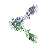
[English] 日本語
Yorodumi
- PDB-3q2v: Crystal structure of mouse E-cadherin ectodomain -

+
Open data


ID or keywords:

Loading...

-
Basic information

Entry
Database: PDB / ID: 3q2v
TitleCrystal structure of mouse E-cadherin ectodomain
ComponentsCadherin-1
KeywordsCELL ADHESION / cadherin / calcium binding
Function / homology
Function and homology information


uterine epithelium development / Apoptotic cleavage of cell adhesion proteins / regulation of branching involved in salivary gland morphogenesis / salivary gland cavitation / regulation of protein catabolic process at postsynapse, modulating synaptic transmission / RHO GTPases activate IQGAPs / Adherens junctions interactions / Degradation of the extracellular matrix / positive regulation of cell-cell adhesion / Integrin cell surface interactions ...uterine epithelium development / Apoptotic cleavage of cell adhesion proteins / regulation of branching involved in salivary gland morphogenesis / salivary gland cavitation / regulation of protein catabolic process at postsynapse, modulating synaptic transmission / RHO GTPases activate IQGAPs / Adherens junctions interactions / Degradation of the extracellular matrix / positive regulation of cell-cell adhesion / Integrin cell surface interactions / lateral loop / regulation of neuron migration / negative regulation of axon extension / cell-cell adhesion mediated by cadherin / regulation of protein localization to cell surface / trophectodermal cell differentiation / alpha-catenin binding / epithelial cell morphogenesis / flotillin complex / Schmidt-Lanterman incisure / bicellular tight junction assembly / calcium-dependent cell-cell adhesion via plasma membrane cell adhesion molecules / intestinal epithelial cell development / node of Ranvier / protein metabolic process / catenin complex / negative regulation of protein processing / cell-cell junction assembly / negative regulation of protein localization to plasma membrane / adherens junction organization / apical junction complex / cochlea development / homophilic cell adhesion via plasma membrane adhesion molecules / microvillus / decidualization / canonical Wnt signaling pathway / establishment of skin barrier / axon terminus / synapse assembly / cytoskeletal protein binding / embryo implantation / protein tyrosine kinase binding / cell periphery / protein localization to plasma membrane / adherens junction / sensory perception of sound / cellular response to amino acid stimulus / negative regulation of canonical Wnt signaling pathway / cell morphogenesis / beta-catenin binding / cell-cell adhesion / negative regulation of epithelial cell proliferation / regulation of protein localization / cell-cell junction / apical part of cell / actin cytoskeleton organization / postsynapse / regulation of gene expression / basolateral plasma membrane / protein phosphatase binding / in utero embryonic development / molecular adaptor activity / endosome / cadherin binding / protein domain specific binding / axon / glutamatergic synapse / calcium ion binding / Golgi apparatus / cell surface / identical protein binding / plasma membrane / cytoplasm
Similarity search - Function
Cadherin prodomain like / Cadherin prodomain / Cadherin prodomain like / Cadherin, Y-type LIR-motif / Cadherin, Y-type LIR-motif / Catenin binding domain superfamily / Cadherins / Cadherin / Cadherin conserved site / Cadherin domain signature. ...Cadherin prodomain like / Cadherin prodomain / Cadherin prodomain like / Cadherin, Y-type LIR-motif / Cadherin, Y-type LIR-motif / Catenin binding domain superfamily / Cadherins / Cadherin / Cadherin conserved site / Cadherin domain signature. / Cadherin repeats. / Cadherin domain / Cadherins domain profile. / Cadherin-like / Cadherin-like superfamily / Immunoglobulin-like / Sandwich / Mainly Beta
Similarity search - Domain/homology
alpha-D-mannopyranose / : / Cadherin-1
Similarity search - Component
Biological speciesMus musculus (house mouse)
MethodX-RAY DIFFRACTION / SYNCHROTRON / MOLECULAR REPLACEMENT / Resolution: 3.4 Å
AuthorsJin, X. / Harrison, O.J. / Shapiro, L.
CitationJournal: Structure / Year: 2011
Title: The extracellular architecture of adherens junctions revealed by crystal structures of type I cadherins.
Authors: Harrison, O.J. / Jin, X. / Hong, S. / Bahna, F. / Ahlsen, G. / Brasch, J. / Wu, Y. / Vendome, J. / Felsovalyi, K. / Hampton, C.M. / Troyanovsky, R.B. / Ben-Shaul, A. / Frank, J. / ...Authors: Harrison, O.J. / Jin, X. / Hong, S. / Bahna, F. / Ahlsen, G. / Brasch, J. / Wu, Y. / Vendome, J. / Felsovalyi, K. / Hampton, C.M. / Troyanovsky, R.B. / Ben-Shaul, A. / Frank, J. / Troyanovsky, S.M. / Shapiro, L. / Honig, B.
History
DepositionDec 20, 2010Deposition site: RCSB / Processing site: RCSB
Revision 1.0Apr 6, 2011Provider: repository / Type: Initial release
Revision 1.1Jul 13, 2011Group: Version format compliance
Revision 1.2Jul 29, 2020Group: Advisory / Data collection ...Advisory / Data collection / Database references / Derived calculations / Structure summary
Category: chem_comp / database_PDB_caveat ...chem_comp / database_PDB_caveat / entity / pdbx_chem_comp_identifier / pdbx_entity_nonpoly / pdbx_struct_conn_angle / struct_conn / struct_conn_type / struct_ref_seq_dif / struct_site / struct_site_gen
Item: _chem_comp.name / _chem_comp.type ..._chem_comp.name / _chem_comp.type / _entity.pdbx_description / _pdbx_entity_nonpoly.name / _pdbx_struct_conn_angle.ptnr1_auth_asym_id / _pdbx_struct_conn_angle.ptnr1_auth_comp_id / _pdbx_struct_conn_angle.ptnr1_auth_seq_id / _pdbx_struct_conn_angle.ptnr1_label_asym_id / _pdbx_struct_conn_angle.ptnr1_label_atom_id / _pdbx_struct_conn_angle.ptnr1_label_comp_id / _pdbx_struct_conn_angle.ptnr1_label_seq_id / _pdbx_struct_conn_angle.ptnr2_auth_asym_id / _pdbx_struct_conn_angle.ptnr2_auth_comp_id / _pdbx_struct_conn_angle.ptnr2_auth_seq_id / _pdbx_struct_conn_angle.ptnr2_label_asym_id / _pdbx_struct_conn_angle.ptnr2_label_atom_id / _pdbx_struct_conn_angle.ptnr2_label_comp_id / _pdbx_struct_conn_angle.ptnr3_auth_asym_id / _pdbx_struct_conn_angle.ptnr3_auth_comp_id / _pdbx_struct_conn_angle.ptnr3_auth_seq_id / _pdbx_struct_conn_angle.ptnr3_label_asym_id / _pdbx_struct_conn_angle.ptnr3_label_atom_id / _pdbx_struct_conn_angle.ptnr3_label_comp_id / _pdbx_struct_conn_angle.ptnr3_label_seq_id / _pdbx_struct_conn_angle.value / _struct_conn.conn_type_id / _struct_conn.id / _struct_conn.pdbx_dist_value / _struct_conn.pdbx_leaving_atom_flag / _struct_conn.pdbx_role / _struct_conn.ptnr1_auth_asym_id / _struct_conn.ptnr1_auth_comp_id / _struct_conn.ptnr1_auth_seq_id / _struct_conn.ptnr1_label_asym_id / _struct_conn.ptnr1_label_atom_id / _struct_conn.ptnr1_label_comp_id / _struct_conn.ptnr1_label_seq_id / _struct_conn.ptnr2_auth_asym_id / _struct_conn.ptnr2_auth_comp_id / _struct_conn.ptnr2_auth_seq_id / _struct_conn.ptnr2_label_asym_id / _struct_conn.ptnr2_label_atom_id / _struct_conn.ptnr2_label_comp_id / _struct_conn_type.id / _struct_ref_seq_dif.details
Description: Carbohydrate remediation / Provider: repository / Type: Remediation

-
Structure visualization

Structure viewerMolecule:
MolmilJmol/JSmol

Downloads & links

-
Assembly

Deposited unit
A: Cadherin-1
B: Cadherin-1
hetero molecules


Theoretical massNumber of molelcules
Total (without water)126,18346
Polymers121,8692
Non-polymers4,31544
Water2,432135
1


  • Idetical with deposited unit
  • defined by author&software
TypeNameSymmetry operationNumber
identity operation1_555x,y,z1
Buried area7020 Å2
ΔGint-94 kcal/mol
Surface area52380 Å2
MethodPISA
Unit cell
Length a, b, c (Å)119.140, 79.697, 176.001
Angle α, β, γ (deg.)90.00, 98.56, 90.00
Int Tables number5
Space group name H-MC121

-
Components

#1: Protein Cadherin-1 / ARC-1 / Epithelial cadherin / E-cadherin / Uvomorulin / E-Cad/CTF1 / E-Cad/CTF2 / E-Cad/CTF3


Mass: 60934.414 Da / Num. of mol.: 2 / Fragment: UNP residues 157-700
Source method: isolated from a genetically manipulated source
Source: (gene. exp.) Mus musculus (house mouse) / Gene: Cdh1 / Cell line (production host): HEK293 / Production host: Homo sapiens (human) / References: UniProt: P09803
#2: Chemical...
ChemComp-CA / CALCIUM ION


Mass: 40.078 Da / Num. of mol.: 24 / Source method: obtained synthetically / Formula: Ca
#3: Chemical ChemComp-MN / MANGANESE (II) ION


Mass: 54.938 Da / Num. of mol.: 2 / Source method: obtained synthetically / Formula: Mn
#4: Sugar
ChemComp-MAN / alpha-D-mannopyranose


Type: D-saccharide, alpha linking / Mass: 180.156 Da / Num. of mol.: 18
Source method: isolated from a genetically manipulated source
Formula: C6H12O6
IdentifierTypeProgram
DManpaCONDENSED IUPAC CARBOHYDRATE SYMBOLGMML 1.0
a-D-mannopyranoseCOMMON NAMEGMML 1.0
a-D-ManpIUPAC CARBOHYDRATE SYMBOLPDB-CARE 1.0
ManSNFG CARBOHYDRATE SYMBOLGMML 1.0
#5: Water ChemComp-HOH / water


Mass: 18.015 Da / Num. of mol.: 135 / Source method: isolated from a natural source / Formula: H2O

-
Experimental details

-
Experiment

ExperimentMethod: X-RAY DIFFRACTION / Number of used crystals: 1

-
Sample preparation

CrystalDensity Matthews: 3.39 Å3/Da / Density % sol: 63.72 %
Crystal growTemperature: 293 K / pH: 8.5
Details: 11% (w/v) PEG 5000 monomethyl ether (MME), 0.1M Bicine pH 8.5, 10mM CaCl2, 30mM MnCl2, 2% (v/v) dioxane, 4% (v/v) 1-butanol , VAPOR DIFFUSION, HANGING DROP, temperature 293K

-
Data collection

DiffractionMean temperature: 100 K
Diffraction sourceSource: SYNCHROTRON / Site: NSLS / Beamline: X4A / Wavelength: 0.9793
DetectorType: ADSC QUANTUM 4 / Detector: CCD / Date: Jul 11, 2008
RadiationMonochromator: SI (111) / Protocol: SINGLE WAVELENGTH / Monochromatic (M) / Laue (L): M / Scattering type: x-ray
Radiation wavelengthWavelength: 0.9793 Å / Relative weight: 1
ReflectionResolution: 3.4→20 Å / Num. obs: 24708 / % possible obs: 99.5 % / Observed criterion σ(I): 1

-
Processing

Software
NameVersionClassification
ADSCQuantumdata collection
MOLREPphasing
PHENIX(phenix.refine: 1.6_289)refinement
DENZOdata reduction
SCALEPACKdata scaling
RefinementMethod to determine structure: MOLECULAR REPLACEMENT / Resolution: 3.4→19.92 Å / SU ML: 0.5 / σ(F): 1.36 / Stereochemistry target values: ML
RfactorNum. reflection% reflection
Rfree0.293 1136 5.04 %
Rwork0.23 --
obs0.233 22536 99.8 %
all-24708 -
Solvent computationShrinkage radii: 0.9 Å / VDW probe radii: 1.11 Å / Solvent model: FLAT BULK SOLVENT MODEL / Bsol: 37.59 Å2 / ksol: 0.26 e/Å3
Displacement parameters
Baniso -1Baniso -2Baniso -3
1--19.8311 Å20 Å2-0.6393 Å2
2--27.6808 Å2-0 Å2
3----7.8497 Å2
Refinement stepCycle: LAST / Resolution: 3.4→19.92 Å
ProteinNucleic acidLigandSolventTotal
Num. atoms7503 0 224 135 7862
Refine LS restraints
Refine-IDTypeDev idealNumber
X-RAY DIFFRACTIONf_bond_d0.0097869
X-RAY DIFFRACTIONf_angle_d1.41610780
X-RAY DIFFRACTIONf_dihedral_angle_d19.1892900
X-RAY DIFFRACTIONf_chiral_restr0.0941305
X-RAY DIFFRACTIONf_plane_restr0.0071399
LS refinement shell
Resolution (Å)Rfactor RfreeNum. reflection RfreeRfactor RworkNum. reflection RworkRefine-ID% reflection obs (%)
3.4-3.55390.37551430.29922632X-RAY DIFFRACTION100
3.5539-3.74010.39741240.26942671X-RAY DIFFRACTION100
3.7401-3.97270.27541240.22612701X-RAY DIFFRACTION100
3.9727-4.27670.25841280.20082648X-RAY DIFFRACTION100
4.2767-4.70190.26861640.19992648X-RAY DIFFRACTION100
4.7019-5.37060.24341510.20282683X-RAY DIFFRACTION100
5.3706-6.72290.28591340.24652695X-RAY DIFFRACTION100
6.7229-19.92460.29241680.22922722X-RAY DIFFRACTION100

+
About Yorodumi

-
News

-
Feb 9, 2022. New format data for meta-information of EMDB entries

New format data for meta-information of EMDB entries

  • Version 3 of the EMDB header file is now the official format.
  • The previous official version 1.9 will be removed from the archive.

Related info.:EMDB header

External links:wwPDB to switch to version 3 of the EMDB data model

-
Aug 12, 2020. Covid-19 info

Covid-19 info

URL: https://pdbjlvh1.pdbj.org/emnavi/covid19.php

New page: Covid-19 featured information page in EM Navigator.

Related info.:Covid-19 info / Mar 5, 2020. Novel coronavirus structure data

+
Mar 5, 2020. Novel coronavirus structure data

Novel coronavirus structure data

Related info.:Yorodumi Speices / Aug 12, 2020. Covid-19 info

External links:COVID-19 featured content - PDBj / Molecule of the Month (242):Coronavirus Proteases

+
Jan 31, 2019. EMDB accession codes are about to change! (news from PDBe EMDB page)

EMDB accession codes are about to change! (news from PDBe EMDB page)

  • The allocation of 4 digits for EMDB accession codes will soon come to an end. Whilst these codes will remain in use, new EMDB accession codes will include an additional digit and will expand incrementally as the available range of codes is exhausted. The current 4-digit format prefixed with “EMD-” (i.e. EMD-XXXX) will advance to a 5-digit format (i.e. EMD-XXXXX), and so on. It is currently estimated that the 4-digit codes will be depleted around Spring 2019, at which point the 5-digit format will come into force.
  • The EM Navigator/Yorodumi systems omit the EMD- prefix.

Related info.:Q: What is EMD? / ID/Accession-code notation in Yorodumi/EM Navigator

External links:EMDB Accession Codes are Changing Soon! / Contact to PDBj

+
Jul 12, 2017. Major update of PDB

Major update of PDB

  • wwPDB released updated PDB data conforming to the new PDBx/mmCIF dictionary.
  • This is a major update changing the version number from 4 to 5, and with Remediation, in which all the entries are updated.
  • In this update, many items about electron microscopy experimental information are reorganized (e.g. em_software).
  • Now, EM Navigator and Yorodumi are based on the updated data.

External links:wwPDB Remediation / Enriched Model Files Conforming to OneDep Data Standards Now Available in the PDB FTP Archive

-
Yorodumi

Thousand views of thousand structures

  • Yorodumi is a browser for structure data from EMDB, PDB, SASBDB, etc.
  • This page is also the successor to EM Navigator detail page, and also detail information page/front-end page for Omokage search.
  • The word "yorodu" (or yorozu) is an old Japanese word meaning "ten thousand". "mi" (miru) is to see.

Related info.:EMDB / PDB / SASBDB / Comparison of 3 databanks / Yorodumi Search / Aug 31, 2016. New EM Navigator & Yorodumi / Yorodumi Papers / Jmol/JSmol / Function and homology information / Changes in new EM Navigator and Yorodumi

Read more