 Movie
Movie Controller

[English] 日本語
 Yorodumi
- PDB-3q1i: Polo-like kinase I Polo-box domain in complex with FMPPPMSpSM pho... -
+ Open data

- Basic information
| Entry | Database: PDB / ID: 3q1i | ||||||
|---|---|---|---|---|---|---|---|
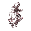
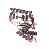
| Title | Polo-like kinase I Polo-box domain in complex with FMPPPMSpSM phosphopeptide from TCERG1 | ||||||
 Components Components |
| ||||||
 Keywords Keywords | TRANSFERASE / KINASE / PEPTIDE BINDING PROTEIN / Plk1 | ||||||
| Function / homology |  Function and homology information Function and homology informationpositive regulation of mitotic nuclear envelope disassembly / Mitotic Telophase/Cytokinesis / regulation of protein localization to cell cortex / Mitotic Metaphase/Anaphase Transition / synaptonemal complex disassembly / Activation of NIMA Kinases NEK9, NEK6, NEK7 / polo kinase / Phosphorylation of Emi1 / homologous chromosome segregation / mitotic nuclear membrane disassembly ...positive regulation of mitotic nuclear envelope disassembly / Mitotic Telophase/Cytokinesis / regulation of protein localization to cell cortex / Mitotic Metaphase/Anaphase Transition / synaptonemal complex disassembly / Activation of NIMA Kinases NEK9, NEK6, NEK7 / polo kinase / Phosphorylation of Emi1 / homologous chromosome segregation / mitotic nuclear membrane disassembly / metaphase/anaphase transition of mitotic cell cycle / female meiosis chromosome segregation / anaphase-promoting complex binding / synaptonemal complex / Phosphorylation of the APC/C / astral microtubule organization / Golgi inheritance / mitotic cleavage furrow formation / outer kinetochore / microtubule bundle formation / mitotic chromosome condensation / double-strand break repair via alternative nonhomologous end joining / regulation of mitotic spindle assembly / Polo-like kinase mediated events / Golgi Cisternae Pericentriolar Stack Reorganization / positive regulation of mitotic metaphase/anaphase transition / RNA polymerase binding / transcription elongation factor activity / centrosome cycle / positive regulation of ubiquitin-dependent protein catabolic process / regulation of mitotic metaphase/anaphase transition / sister chromatid cohesion / mitotic spindle pole / mitotic spindle assembly checkpoint signaling / ubiquitin-like protein conjugating enzyme binding / regulation of mitotic cell cycle phase transition / spindle midzone / regulation of anaphase-promoting complex-dependent catabolic process / mitotic G2 DNA damage checkpoint signaling / mitotic cytokinesis / mitotic sister chromatid segregation / negative regulation of transcription elongation by RNA polymerase II / negative regulation of double-strand break repair via homologous recombination / Regulation of MITF-M-dependent genes involved in cell cycle and proliferation / Cyclin A/B1/B2 associated events during G2/M transition / protein localization to chromatin / Loss of Nlp from mitotic centrosomes / Loss of proteins required for interphase microtubule organization from the centrosome / Amplification of signal from unattached kinetochores via a MAD2 inhibitory signal / mRNA Splicing - Major Pathway / Recruitment of mitotic centrosome proteins and complexes / Recruitment of NuMA to mitotic centrosomes / Anchoring of the basal body to the plasma membrane / regulation of mitotic cell cycle / Mitotic Prometaphase / EML4 and NUDC in mitotic spindle formation / RNA splicing / AURKA Activation by TPX2 / Resolution of Sister Chromatid Cohesion / mitotic spindle organization / Condensation of Prophase Chromosomes / regulation of cytokinesis / transcription coregulator activity / establishment of protein localization / centriole / positive regulation of transcription elongation by RNA polymerase II / RHO GTPases Activate Formins / APC/C:Cdh1 mediated degradation of Cdc20 and other APC/C:Cdh1 targeted proteins in late mitosis/early G1 / positive regulation of protein localization to nucleus / kinetochore / G2/M transition of mitotic cell cycle / spindle / centriolar satellite / mRNA processing / spindle pole / The role of GTSE1 in G2/M progression after G2 checkpoint / transcription corepressor activity / Separation of Sister Chromatids / Regulation of PLK1 Activity at G2/M Transition / double-strand break repair / mitotic cell cycle / positive regulation of proteasomal ubiquitin-dependent protein catabolic process / microtubule cytoskeleton / midbody / microtubule binding / protein phosphorylation / RNA polymerase II-specific DNA-binding transcription factor binding / protein kinase activity / transcription coactivator activity / regulation of cell cycle / nuclear speck / protein serine kinase activity / protein serine/threonine kinase activity / centrosome / protein kinase binding / negative regulation of apoptotic process / chromatin / magnesium ion binding / negative regulation of transcription by RNA polymerase II / positive regulation of transcription by RNA polymerase II Similarity search - Function | ||||||
| Biological species |  Homo sapiens (human) Homo sapiens (human) | ||||||
| Method |  X-RAY DIFFRACTION / SYNCHROTRON / MOLECULAR REPLACEMENT / Resolution: 1.4 Å X-RAY DIFFRACTION / SYNCHROTRON / MOLECULAR REPLACEMENT / Resolution: 1.4 Å | ||||||
 Authors Authors | Sledz, P. / Hyvonen, M. / Abell, C. | ||||||
 Citation Citation | Journal: Angew.Chem.Int.Ed.Engl. / Year: 2011 Title: From crystal packing to molecular recognition: prediction and discovery of a binding site on the surface of polo-like kinase 1 Authors: Sledz, P. / Stubbs, C.J. / Lang, S. / Yang, Y.Q. / McKenzie, G.J. / Venkitaraman, A.R. / Hyvonen, M. / Abell, C. | ||||||
| History |
|
- Structure visualization
| Structure viewer | Molecule: MolmilJmol/JSmol |
|---|
- Downloads & links
-Download
| PDBx/mmCIF format | 3q1i.cif.gz | 99.9 KB | Display | PDBx/mmCIF format |
|---|---|---|---|---|
| PDB format | pdb3q1i.ent.gz | 74.9 KB | Display | PDB format |
| PDBx/mmJSON format | 3q1i.json.gz | Tree view | PDBx/mmJSON format | |
| Others |  Other downloads Other downloads |
-Validation report
| Arichive directory | https://data.pdbj.org/pub/pdb/validation_reports/q1/3q1iftp://data.pdbj.org/pub/pdb/validation_reports/q1/3q1i | HTTPS FTP |
|---|
-Related structure data
| Related structure data |  3p2wC 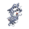 3p2zC  3p34C  3p35C 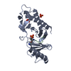 3p36C  3p37C  1umwS S: Starting model for refinement C: citing same article ( |
|---|---|
| Similar structure data |


-Links
 PDBj
PDBj


- Assembly
| Deposited unit | 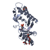
| ||||||||
|---|---|---|---|---|---|---|---|---|---|
| 1 |
| ||||||||
| Unit cell |
|
-Components
-Protein / Protein/peptide , 2 types, 2 molecules AE
| #1: Protein | Mass: 26755.518 Da / Num. of mol.: 1 / Fragment: Polo-box domain, residues 371-594 Source method: isolated from a genetically manipulated source Source: (gene. exp.) Homo sapiens (human) / Gene: PLK1, PLK / Production host:  |
|---|---|
| #2: Protein/peptide | Mass: 1128.301 Da / Num. of mol.: 1 / Source method: obtained synthetically / Details: Synthetic phosphopeptide / References: UniProt: O14776 |
-Non-polymers , 4 types, 195 molecules 


| #3: Chemical | ChemComp-1PE /  Mass: 238.278 Da / Num. of mol.: 1 / Source method: obtained synthetically / Formula: C10H22O6 / Comment: precipitant*YM Mass: 238.278 Da / Num. of mol.: 1 / Source method: obtained synthetically / Formula: C10H22O6 / Comment: precipitant*YM |
|---|---|
| #4: Chemical | ChemComp-PEG /  Mass: 106.120 Da / Num. of mol.: 1 / Source method: obtained synthetically / Formula: C4H10O3 Mass: 106.120 Da / Num. of mol.: 1 / Source method: obtained synthetically / Formula: C4H10O3 |
| #5: Chemical | ChemComp-PG0 /  Mass: 120.147 Da / Num. of mol.: 1 / Source method: obtained synthetically / Formula: C5H12O3 / Comment: inhibitor, precipitant*YM Mass: 120.147 Da / Num. of mol.: 1 / Source method: obtained synthetically / Formula: C5H12O3 / Comment: inhibitor, precipitant*YM |
| #6: Water | ChemComp-HOH / Mass: 18.015 Da / Num. of mol.: 192 / Source method: isolated from a natural source / Formula: H2O |
-Details
| Has protein modification | Y |
|---|
-Experimental details
-Experiment
| Experiment | Method: X-RAY DIFFRACTION / Number of used crystals: 1 |
|---|
- Sample preparation
| Crystal | Density Matthews: 1.85 Å3/Da / Density % sol: 33.64 % |
|---|---|
| Crystal grow | Temperature: 293 K / Method: vapor diffusion, sitting drop / pH: 6.5 Details: 30% PEG400, 0.1M MES, pH 6.5, VAPOR DIFFUSION, SITTING DROP, temperature 293K |
-Data collection
| Diffraction | Mean temperature: 100 K |
|---|---|
| Diffraction source | Source: SYNCHROTRON / Site: SOLEIL  / Beamline: PROXIMA 1 / Wavelength: 0.9184 Å / Beamline: PROXIMA 1 / Wavelength: 0.9184 Å |
| Detector | Date: Nov 17, 2010 |
| Radiation | Protocol: SINGLE WAVELENGTH / Monochromatic (M) / Laue (L): M / Scattering type: x-ray |
| Radiation wavelength | Wavelength: 0.9184 Å / Relative weight: 1 |
| Reflection | Resolution: 1.4→56.54 Å / Num. all: 40284 / Num. obs: 38959 / % possible obs: 97 % |
- Processing
| Software |
| ||||||||||||||||||||||||||||||||||||||||||||||||||||||||||||||||||||||||||||||||||||||||||||||||||||||||||||||||||||||||||||||||||||||||||||||||||||||||||||||||||||||||||
|---|---|---|---|---|---|---|---|---|---|---|---|---|---|---|---|---|---|---|---|---|---|---|---|---|---|---|---|---|---|---|---|---|---|---|---|---|---|---|---|---|---|---|---|---|---|---|---|---|---|---|---|---|---|---|---|---|---|---|---|---|---|---|---|---|---|---|---|---|---|---|---|---|---|---|---|---|---|---|---|---|---|---|---|---|---|---|---|---|---|---|---|---|---|---|---|---|---|---|---|---|---|---|---|---|---|---|---|---|---|---|---|---|---|---|---|---|---|---|---|---|---|---|---|---|---|---|---|---|---|---|---|---|---|---|---|---|---|---|---|---|---|---|---|---|---|---|---|---|---|---|---|---|---|---|---|---|---|---|---|---|---|---|---|---|---|---|---|---|---|---|---|
| Refinement | Method to determine structure: MOLECULAR REPLACEMENT Starting model: 1UMW chain A Resolution: 1.4→56.54 Å / Cor.coef. Fo:Fc: 0.959 / Cor.coef. Fo:Fc free: 0.949 / SU B: 1.994 / SU ML: 0.046 / Cross valid method: THROUGHOUT / ESU R: 0.075 / ESU R Free: 0.075 / Stereochemistry target values: MAXIMUM LIKELIHOOD
| ||||||||||||||||||||||||||||||||||||||||||||||||||||||||||||||||||||||||||||||||||||||||||||||||||||||||||||||||||||||||||||||||||||||||||||||||||||||||||||||||||||||||||
| Solvent computation | Ion probe radii: 0.8 Å / Shrinkage radii: 0.8 Å / VDW probe radii: 1.4 Å / Solvent model: MASK | ||||||||||||||||||||||||||||||||||||||||||||||||||||||||||||||||||||||||||||||||||||||||||||||||||||||||||||||||||||||||||||||||||||||||||||||||||||||||||||||||||||||||||
| Displacement parameters | Biso mean: 17.809 Å2
| ||||||||||||||||||||||||||||||||||||||||||||||||||||||||||||||||||||||||||||||||||||||||||||||||||||||||||||||||||||||||||||||||||||||||||||||||||||||||||||||||||||||||||
| Refinement step | Cycle: LAST / Resolution: 1.4→56.54 Å
| ||||||||||||||||||||||||||||||||||||||||||||||||||||||||||||||||||||||||||||||||||||||||||||||||||||||||||||||||||||||||||||||||||||||||||||||||||||||||||||||||||||||||||
| Refine LS restraints |
| ||||||||||||||||||||||||||||||||||||||||||||||||||||||||||||||||||||||||||||||||||||||||||||||||||||||||||||||||||||||||||||||||||||||||||||||||||||||||||||||||||||||||||
| LS refinement shell | Resolution: 1.4→1.436 Å / Total num. of bins used: 20
| ||||||||||||||||||||||||||||||||||||||||||||||||||||||||||||||||||||||||||||||||||||||||||||||||||||||||||||||||||||||||||||||||||||||||||||||||||||||||||||||||||||||||||
| Refinement TLS params. | Method: refined / Origin x: 8.095 Å / Origin y: -0.323 Å / Origin z: 14.42 Å
|
