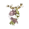
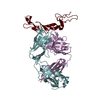
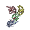
- PDB-3pvl: Structure of myosin VIIa MyTH4-FERM-SH3 in complex with the CEN1 ... -
+
データを開く
IDまたはキーワード:
読み込み中...
-
基本情報
登録情報
データベース: PDB / ID: 3pvl
タイトル
Structure of myosin VIIa MyTH4-FERM-SH3 in complex with the CEN1 of Sans
要素
Myosin VIIa isoform 1
Usher syndrome type-1G protein
キーワード
MOTOR PROTEIN/PROTEIN TRANSPORT (機関 (機械)) / protein complex (タンパク質複合体) / Novel folding / protein cargo binding / cargo proteins (貨物) / MOTOR PROTEIN-PROTEIN TRANSPORT complex (機関 (機械))
機能・相同性
機能・相同性情報
: / photoreceptor cell cilium / pigment granule localization / upper tip-link density / pigment granule transport / The canonical retinoid cycle in rods (twilight vision) / microfilament motor activity => GO:0000146 / stereocilium base / myosin VII complex / phagolysosome assembly ...: / photoreceptor cell cilium / pigment granule localization / upper tip-link density / pigment granule transport / The canonical retinoid cycle in rods (twilight vision) / microfilament motor activity => GO:0000146 / stereocilium base / myosin VII complex / phagolysosome assembly / inner ear receptor cell differentiation / protein localization => GO:0008104 / regulation of clathrin-dependent endocytosis / 平衡感覚 / sensory perception of light stimulus / mechanoreceptor differentiation / photoreceptor connecting cilium / inner ear receptor cell stereocilium organization / sensory organ development / actin filament-based movement / : / inner ear auditory receptor cell differentiation / 知覚 / stereocilium / cell projection organization / vesicle transport along actin filament / photoreceptor cell maintenance / auditory receptor cell stereocilium organization / Sensory processing of sound by outer hair cells of the cochlea / Sensory processing of sound by inner hair cells of the cochlea / myosin complex / ciliary base / inner ear morphogenesis / microfilament motor activity / spectrin binding / lysosome organization / cochlea development / 微絨毛 / cytoskeletal motor activity / inner ear development / photoreceptor outer segment / カハール体 / 食作用 / 視覚 / photoreceptor inner segment / ciliary basal body / actin filament organization / sensory perception of sound / intracellular protein transport / protein localization / ADP binding / actin filament binding / メラノソーム / マイクロフィラメント / actin binding / 細胞皮質 / vesicle / calmodulin binding / nuclear speck / apical plasma membrane / lysosomal membrane / protein domain specific binding / 中心体 / シナプス / protein-containing complex binding / ATP binding / identical protein binding / 細胞膜 / 細胞質基質 / 細胞質 類似検索 - 分子機能
USH1G, SAM domain / MyTH4 domain / Class VII myosin, motor domain / Myosin VII, FERM domain C-lobe, repeat 1 / Myosin VII, FERM domain C-lobe, repeat 2 / MyTH4 domain / MyTH4 domain superfamily / MyTH4 domain / MyTH4 domain profile. / Domain in Myosin and Kinesin Tails ...USH1G, SAM domain / MyTH4 domain / Class VII myosin, motor domain / Myosin VII, FERM domain C-lobe, repeat 1 / Myosin VII, FERM domain C-lobe, repeat 2 / MyTH4 domain / MyTH4 domain superfamily / MyTH4 domain / MyTH4 domain profile. / Domain in Myosin and Kinesin Tails / Acyl-CoA Binding Protein - #10 / Acyl-CoA Binding Protein / IQ calmodulin-binding motif / Myosin S1 fragment, N-terminal / SAM domain (Sterile alpha motif) / FERM central domain / FERM/acyl-CoA-binding protein superfamily / Short calmodulin-binding motif containing conserved Ile and Gln residues. / Myosin head, motor domain / Myosin head (motor domain) / Myosin motor domain profile. / Myosin. Large ATPases. / IQ motif profile. / IQ motif, EF-hand binding site / Pleckstrin-homology domain (PH domain)/Phosphotyrosine-binding domain (PTB) / FERM central domain / SH3 Domains / PH-domain like / FERM superfamily, second domain / FERM domain / FERM domain profile. / Band 4.1 domain / Band 4.1 homologues / Sterile alpha motif. / Sterile alpha motif domain / Kinesin motor domain superfamily / Sterile alpha motif/pointed domain superfamily / Phosphatidylinositol 3-kinase Catalytic Subunit; Chain A, domain 1 / Ankyrin repeats (3 copies) / Ankyrin repeat profile. / Ankyrin repeat region circular profile. / ankyrin repeats / Ankyrin repeat / Ankyrin repeat-containing domain superfamily / Serine Threonine Protein Phosphatase 5, Tetratricopeptide repeat / SH3 type barrels. / Src homology 3 domains / Ubiquitin-like (UB roll) / SH3-like domain superfamily / Src homology 3 (SH3) domain profile. / SH3ドメイン / Αソレノイド / PH-like domain superfamily / Ubiquitin-like domain superfamily / Roll / Roll / Up-down Bundle / P-loop containing nucleoside triphosphate hydrolase / Mainly Beta / Mainly Alpha / Alpha Beta 類似検索 - ドメイン・相同性
 ムービー
ムービー コントローラー


 データを開く
 基本情報
基本情報 要素
要素 キーワード
キーワード MOTOR PROTEIN/PROTEIN TRANSPORT (機関 (機械)) /
  機能・相同性情報

 データ登録者
 引用
引用 構造の表示
構造の表示 ダウンロードとリンク
 その他のダウンロード


 PDBj
PDBj


 集合体
集合体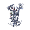


 分子量: 92.094 Da / 分子数: 9 / 由来タイプ: 合成 / 式: C3H8O3
 分子量: 94.971 Da / 分子数: 2 / 由来タイプ: 合成 / 式: PO4
 分子量: 18.015 Da / 分子数: 38 / 由来タイプ: 天然 / 式: H2O
 試料調製
試料調製 / ビームライン: BL17U / 波長: 1.5418 Å
 解析
解析