 Movie
Movie Controller


+ Open data

- Basic information
| Entry | Database: PDB / ID: 3pv3 | ||||||
|---|---|---|---|---|---|---|---|
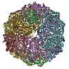
| Title | Structure of Legionella fallonii DegQ (S193A variant) | ||||||
 Components Components |
| ||||||
 Keywords Keywords | HYDROLASE / trypsin fold / PDZ domain / chaperone protease | ||||||
| Function / homology |  Function and homology information Function and homology information | ||||||
| Biological species |  Legionella fallonii (bacteria) Legionella fallonii (bacteria) | ||||||
| Method |  X-RAY DIFFRACTION / SYNCHROTRON / MOLECULAR REPLACEMENT / Resolution: 3.1 Å X-RAY DIFFRACTION / SYNCHROTRON / MOLECULAR REPLACEMENT / Resolution: 3.1 Å | ||||||
 Authors Authors | Wrase, R. / Scott, H. / Hilgenfeld, R. / Hansen, G. | ||||||
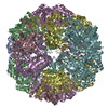
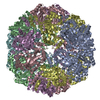
 Citation Citation | Journal: Proc.Natl.Acad.Sci.USA / Year: 2011 Title: The Legionella HtrA homologue DegQ is a self-compartmentizing protease that forms large 12-meric assemblies. Authors: Wrase, R. / Scott, H. / Hilgenfeld, R. / Hansen, G. | ||||||
| History |
|
- Structure visualization
| Structure viewer | Molecule: MolmilJmol/JSmol |
|---|
- Downloads & links
-Download
| PDBx/mmCIF format | 3pv3.cif.gz | 294.2 KB | Display | PDBx/mmCIF format |
|---|---|---|---|---|
| PDB format | pdb3pv3.ent.gz | 237.8 KB | Display | PDB format |
| PDBx/mmJSON format | 3pv3.json.gz | Tree view | PDBx/mmJSON format | |
| Others |  Other downloads Other downloads |
-Validation report
| Arichive directory | https://data.pdbj.org/pub/pdb/validation_reports/pv/3pv3ftp://data.pdbj.org/pub/pdb/validation_reports/pv/3pv3 | HTTPS FTP |
|---|
-Related structure data
| Related structure data |  3pv2SC 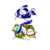 3pv4C  3pv5C S: Starting model for refinement C: citing same article ( |
|---|---|
| Similar structure data |


-Links
 PDBj
PDBj
- Assembly
| Deposited unit | 
| ||||||||
|---|---|---|---|---|---|---|---|---|---|
| 1 | 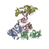
| ||||||||
| Unit cell |
|
-Components
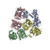
| #1: Protein | Mass: 48016.637 Da / Num. of mol.: 4 / Mutation: S193A Source method: isolated from a genetically manipulated source Source: (gene. exp.) Legionella fallonii (bacteria) / Plasmid: pQE-30 / Production host: #2: Protein/peptide | Mass: 1720.111 Da / Num. of mol.: 4 / Source method: isolated from a natural source / Source: (natural) Sequence details | A MIXTURE OF NATURAL OCCURRING PEPTIDE FRAGMENTS WERE FOUND IN THE ACTIVE SITE FOR WHICH THE ...A MIXTURE OF NATURAL OCCURRING PEPTIDE FRAGMENTS WERE FOUND IN THE ACTIVE SITE FOR WHICH THE SEQUENCE IS UNKNOWN. | |
|---|
-Experimental details
-Experiment
| Experiment | Method: X-RAY DIFFRACTION / Number of used crystals: 1 |
|---|
- Sample preparation
| Crystal | Density Matthews: 3.03 Å3/Da / Density % sol: 59.4 % |
|---|---|
| Crystal grow | Temperature: 292 K / Method: vapor diffusion, sitting drop / pH: 9.5 Details: 22.5% (v/v) PEG 400, 100 mM glycine pH 9.5, vapor diffusion, sitting drop, temperature 292K |
-Data collection
| Diffraction | Mean temperature: 100 K | |||||||||||||||||||||||||||||||||||||||||||||||||||||||||||||||||||||||||||||
|---|---|---|---|---|---|---|---|---|---|---|---|---|---|---|---|---|---|---|---|---|---|---|---|---|---|---|---|---|---|---|---|---|---|---|---|---|---|---|---|---|---|---|---|---|---|---|---|---|---|---|---|---|---|---|---|---|---|---|---|---|---|---|---|---|---|---|---|---|---|---|---|---|---|---|---|---|---|---|
| Diffraction source | Source: SYNCHROTRON / Site: BESSY  / Beamline: 14.1 / Wavelength: 0.91841 Å / Beamline: 14.1 / Wavelength: 0.91841 Å | |||||||||||||||||||||||||||||||||||||||||||||||||||||||||||||||||||||||||||||
| Detector | Type: Rayonics MX-225 3x3 CCD / Detector: CCD / Date: Mar 12, 2010 | |||||||||||||||||||||||||||||||||||||||||||||||||||||||||||||||||||||||||||||
| Radiation | Monochromator: Double crystal monochromator, Si(111) / Protocol: SINGLE WAVELENGTH / Monochromatic (M) / Laue (L): M / Scattering type: x-ray | |||||||||||||||||||||||||||||||||||||||||||||||||||||||||||||||||||||||||||||
| Radiation wavelength | Wavelength: 0.91841 Å / Relative weight: 1 | |||||||||||||||||||||||||||||||||||||||||||||||||||||||||||||||||||||||||||||
| Reflection | Resolution: 3.1→119.351 Å / Num. all: 41803 / Num. obs: 41803 / % possible obs: 98.7 % / Redundancy: 2.9 % / Rsym value: 0.095 / Net I/σ(I): 6.6 | |||||||||||||||||||||||||||||||||||||||||||||||||||||||||||||||||||||||||||||
| Reflection shell |
|
- Processing
| Software |
| ||||||||||||||||||||||||||||||||||||||||||||||||||||||||||||||||||||||||||||||||||||||||||||||||||||||||||||||||||||||||||||||||||||||||||||||||||||||||||||||||||||||||||
|---|---|---|---|---|---|---|---|---|---|---|---|---|---|---|---|---|---|---|---|---|---|---|---|---|---|---|---|---|---|---|---|---|---|---|---|---|---|---|---|---|---|---|---|---|---|---|---|---|---|---|---|---|---|---|---|---|---|---|---|---|---|---|---|---|---|---|---|---|---|---|---|---|---|---|---|---|---|---|---|---|---|---|---|---|---|---|---|---|---|---|---|---|---|---|---|---|---|---|---|---|---|---|---|---|---|---|---|---|---|---|---|---|---|---|---|---|---|---|---|---|---|---|---|---|---|---|---|---|---|---|---|---|---|---|---|---|---|---|---|---|---|---|---|---|---|---|---|---|---|---|---|---|---|---|---|---|---|---|---|---|---|---|---|---|---|---|---|---|---|---|---|
| Refinement | Method to determine structure: MOLECULAR REPLACEMENT Starting model: PDB ENTRY 3PV2 Resolution: 3.1→59.61 Å / Cor.coef. Fo:Fc: 0.923 / Cor.coef. Fo:Fc free: 0.884 / Occupancy max: 1 / Occupancy min: 1 / Cross valid method: THROUGHOUT / ESU R Free: 0.504 / Stereochemistry target values: MAXIMUM LIKELIHOOD / Details: HYDROGENS HAVE BEEN ADDED IN THE RIDING POSITIONS
| ||||||||||||||||||||||||||||||||||||||||||||||||||||||||||||||||||||||||||||||||||||||||||||||||||||||||||||||||||||||||||||||||||||||||||||||||||||||||||||||||||||||||||
| Solvent computation | Ion probe radii: 0.8 Å / Shrinkage radii: 0.8 Å / VDW probe radii: 1.4 Å / Solvent model: MASK | ||||||||||||||||||||||||||||||||||||||||||||||||||||||||||||||||||||||||||||||||||||||||||||||||||||||||||||||||||||||||||||||||||||||||||||||||||||||||||||||||||||||||||
| Displacement parameters | Biso mean: 72.179 Å2
| ||||||||||||||||||||||||||||||||||||||||||||||||||||||||||||||||||||||||||||||||||||||||||||||||||||||||||||||||||||||||||||||||||||||||||||||||||||||||||||||||||||||||||
| Refinement step | Cycle: LAST / Resolution: 3.1→59.61 Å
| ||||||||||||||||||||||||||||||||||||||||||||||||||||||||||||||||||||||||||||||||||||||||||||||||||||||||||||||||||||||||||||||||||||||||||||||||||||||||||||||||||||||||||
| Refine LS restraints |
| ||||||||||||||||||||||||||||||||||||||||||||||||||||||||||||||||||||||||||||||||||||||||||||||||||||||||||||||||||||||||||||||||||||||||||||||||||||||||||||||||||||||||||
| LS refinement shell | Resolution: 3.1→3.18 Å / Total num. of bins used: 20
|
