 Movie
Movie Controller

[English] 日本語
 Yorodumi
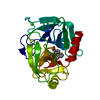
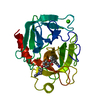
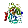
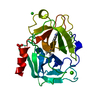
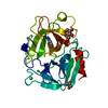
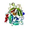
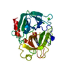
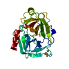
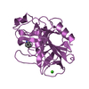
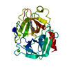
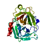
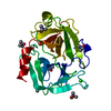
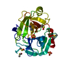
- PDB-3ptb: THE GEOMETRY OF THE REACTIVE SITE AND OF THE PEPTIDE GROUPS IN TR... -
+ Open data

- Basic information
| Entry | Database: PDB / ID: 3ptb | |||||||||
|---|---|---|---|---|---|---|---|---|---|---|
| Title | THE GEOMETRY OF THE REACTIVE SITE AND OF THE PEPTIDE GROUPS IN TRYPSIN, TRYPSINOGEN AND ITS COMPLEXES WITH INHIBITORS | |||||||||
 Components Components | BETA-TRYPSIN | |||||||||
 Keywords Keywords | HYDROLASE (SERINE PROTEINASE) | |||||||||
| Function / homology |  Function and homology information Function and homology informationtrypsin / serpin family protein binding / serine protease inhibitor complex / digestion / endopeptidase activity / serine-type endopeptidase activity / proteolysis / extracellular space / metal ion binding Similarity search - Function | |||||||||
| Biological species |  | |||||||||
| Method |  X-RAY DIFFRACTION / Resolution: 1.7 Å X-RAY DIFFRACTION / Resolution: 1.7 Å | |||||||||
 Authors Authors | Bode, W. / Schwager, P. / Walter, J. | |||||||||
 Citation Citation | Journal: Acta Crystallogr.,Sect.B / Year: 1983 Title: The Geometry of the Reactive Site and of the Peptide Groups in Trypsin, Trypsinogen and its Complexes with Inhibitors Authors: Marquart, M. / Walter, J. / Deisenhofer, J. / Bode, W. / Huber, R. #1: Journal: Miami Winter Symp. / Year: 1976Title: Structural Studies on the Pancreatic Trypsin Inhibitor-Trypsin Complex and its Free Components. Structure and Function Relationships in Serine Protease Inhibition and Catalysis Authors: Bode, W. / Schwager, P. / Huber, R. #2: Journal: J.Mol.Biol. / Year: 1975Title: The Refined Crystal Structure of Bovine Beta-Trypsin at 1.8 Angstroms Resolution. I. Crystallization, Data Collection and Application of Patterson Search Techniques Authors: Fehlhammer, H. / Bode, W. #3: Journal: J.Mol.Biol. / Year: 1975Title: The Refined Crystal Structure of Bovine Beta-Trypsin at 1.8 Angstroms Resolution. II. Crystallographic Refinement, Calcium Binding Site, Benzamidine Binding Site and Active Site at Ph 7.0 Authors: Bode, W. / Schwager, P. #4: Journal: FEBS Lett. / Year: 1975Title: The Single Calcium-Binding Site of Crystalline Bovine Beta-Trypsin Authors: Bode, W. / Schwager, P. | |||||||||
| History |
|
- Structure visualization
| Structure viewer | Molecule: MolmilJmol/JSmol |
|---|
- Downloads & links
-Download
| PDBx/mmCIF format | 3ptb.cif.gz | 53 KB | Display | PDBx/mmCIF format |
|---|---|---|---|---|
| PDB format | pdb3ptb.ent.gz | 37.8 KB | Display | PDB format |
| PDBx/mmJSON format | 3ptb.json.gz | Tree view | PDBx/mmJSON format | |
| Others |  Other downloads Other downloads |
-Validation report
| Summary document | 3ptb_validation.pdf.gz | 431.7 KB | Display | wwPDB validaton report |
|---|---|---|---|---|
| Full document | 3ptb_full_validation.pdf.gz | 434.4 KB | Display | |
| Data in XML | 3ptb_validation.xml.gz | 11.2 KB | Display | |
| Data in CIF | 3ptb_validation.cif.gz | 14.9 KB | Display | |
| Arichive directory | https://data.pdbj.org/pub/pdb/validation_reports/pt/3ptbftp://data.pdbj.org/pub/pdb/validation_reports/pt/3ptb | HTTPS FTP |
-Related structure data
| Similar structure data |
|---|


-Links
 PDBj
PDBj


- Assembly
| Deposited unit | 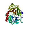
| ||||||||
|---|---|---|---|---|---|---|---|---|---|
| 1 |
| ||||||||
| Unit cell |
| ||||||||
| Atom site foot note | 1: SEE REMARK 4. |
-Components
| #1: Protein | Mass: 23324.287 Da / Num. of mol.: 1 Source method: isolated from a genetically manipulated source Source: (gene. exp.) |
|---|---|
| #2: Chemical | ChemComp-CA /   Mass: 40.078 Da / Num. of mol.: 1 / Source method: obtained synthetically / Formula: Ca Mass: 40.078 Da / Num. of mol.: 1 / Source method: obtained synthetically / Formula: Ca |
| #3: Chemical | ChemComp-BEN /   Mass: 120.152 Da / Num. of mol.: 1 / Source method: obtained synthetically / Formula: C7H8N2 Mass: 120.152 Da / Num. of mol.: 1 / Source method: obtained synthetically / Formula: C7H8N2 |
| #4: Water | ChemComp-HOH /  Mass: 18.015 Da / Num. of mol.: 62 / Source method: isolated from a natural source / Formula: H2O Mass: 18.015 Da / Num. of mol.: 62 / Source method: isolated from a natural source / Formula: H2O |
| Has protein modification | Y |
-Experimental details
-Experiment
| Experiment | Method: X-RAY DIFFRACTION |
|---|
- Sample preparation
| Crystal | Density Matthews: 2.33 Å3/Da / Density % sol: 47.16 % |
|---|---|
| Crystal grow | *PLUS Method: unknown |
- Processing
| Refinement | Resolution: 1.7→7 Å / Rfactor Rwork: 0.182 | ||||||||||||
|---|---|---|---|---|---|---|---|---|---|---|---|---|---|
| Refinement step | Cycle: LAST / Resolution: 1.7→7 Å
|
