 Movie
Movie Controller

[English] 日本語
 Yorodumi
- PDB-3ppl: Crystal structure of an aspartate transaminase (NCgl0237, Cgl0240... -
+ Open data

- Basic information
| Entry | Database: PDB / ID: 3ppl | ||||||
|---|---|---|---|---|---|---|---|
| Title | Crystal structure of an aspartate transaminase (NCgl0237, Cgl0240) from CORYNEBACTERIUM GLUTAMICUM ATCC 13032 KITASATO at 1.25 A resolution | ||||||
 Components Components | ASPARTATE AMINOTRANSFERASE | ||||||
 Keywords Keywords | TRANSFERASE / AMINOTRANSFERASE / DIMER / PLP-DEPENDENT TRANSFERASE-LIKE FOLD / STRUCTURAL GENOMICS / JOINT CENTER FOR STRUCTURAL GENOMICS / JCSG / PROTEIN STRUCTURE INITIATIVE / PSI-BIOLOGY | ||||||
| Function / homology |  Function and homology information Function and homology informationaspartate transaminase / L-aspartate:2-oxoglutarate transaminase activity Similarity search - Function | ||||||
| Biological species |  Corynebacterium glutamicum (bacteria) Corynebacterium glutamicum (bacteria) | ||||||
| Method |  X-RAY DIFFRACTION / SYNCHROTRON / MAD / Resolution: 1.25 Å X-RAY DIFFRACTION / SYNCHROTRON / MAD / Resolution: 1.25 Å | ||||||
 Authors Authors | Joint Center for Structural Genomics (JCSG) | ||||||
 Citation Citation | Journal: To be published Title: Crystal structure of an aspartate transaminase (NCgl0237, Cgl0240) from CORYNEBACTERIUM GLUTAMICUM ATCC 13032 KITASATO at 1.25 A resolution Authors: Joint Center for Structural Genomics (JCSG) | ||||||
| History |
|
- Structure visualization
| Structure viewer | Molecule: MolmilJmol/JSmol |
|---|
- Downloads & links
-Download
| PDBx/mmCIF format | 3ppl.cif.gz | 403.3 KB | Display | PDBx/mmCIF format |
|---|---|---|---|---|
| PDB format | pdb3ppl.ent.gz | 335.5 KB | Display | PDB format |
| PDBx/mmJSON format | 3ppl.json.gz | Tree view | PDBx/mmJSON format | |
| Others |  Other downloads Other downloads |
-Validation report
| Arichive directory | https://data.pdbj.org/pub/pdb/validation_reports/pp/3pplftp://data.pdbj.org/pub/pdb/validation_reports/pp/3ppl | HTTPS FTP |
|---|
-Related structure data
| Similar structure data | |
|---|---|
| Other databases |


-Links
 PDBj
PDBj

- Assembly
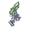
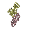
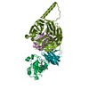
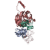
| Deposited unit | 
| ||||||||
|---|---|---|---|---|---|---|---|---|---|
| 1 |
| ||||||||
| Unit cell |
| ||||||||
| Details | ANALYTICAL SIZE EXCLUSION CHROMATOGRAPHY WITH STATIC LIGHT SCATTERING SUPPORTS THE ASSIGNMENT OF A DIMER AS A SIGNIFICANT OLIGOMERIZATION STATE IN SOLUTION. |
-Components
| #1: Protein | Mass: 47044.512 Da / Num. of mol.: 2 Source method: isolated from a genetically manipulated source Source: (gene. exp.) Corynebacterium glutamicum (bacteria) / Strain: ATCC 13032 KITASATO / Gene: aspB, cg0294 / Plasmid: SpeedET / Production host: References: UniProt: Q6M8B5, UniProt: Q8NTR2*PLUS, aspartate transaminase #2: Chemical |   Mass: 247.142 Da / Num. of mol.: 2 / Source method: obtained synthetically / Formula: C8H10NO6P Mass: 247.142 Da / Num. of mol.: 2 / Source method: obtained synthetically / Formula: C8H10NO6P#3: Chemical | Num. of mol.: 2 / Source method: obtained synthetically #4: Chemical |   Mass: 92.094 Da / Num. of mol.: 2 / Source method: obtained synthetically / Formula: C3H8O3 Mass: 92.094 Da / Num. of mol.: 2 / Source method: obtained synthetically / Formula: C3H8O3#5: Water | ChemComp-HOH / |  Mass: 18.015 Da / Num. of mol.: 1228 / Source method: isolated from a natural source / Formula: H2O Mass: 18.015 Da / Num. of mol.: 1228 / Source method: isolated from a natural source / Formula: H2OSequence details | THIS CONSTRUCT WAS EXPRESSED WITH A PURIFICATION TAG MGSDKIHHHHHHENLYFQG. THE TAG WAS REMOVED WITH ...THIS CONSTRUCT WAS EXPRESSED WITH A PURIFICATI | |
|---|
-Experimental details
-Experiment
| Experiment | Method: X-RAY DIFFRACTION / Number of used crystals: 1 |
|---|
- Sample preparation
| Crystal | Density Matthews: 2.44 Å3/Da / Density % sol: 49.63 % |
|---|---|
| Crystal grow | Temperature: 293 K / Method: vapor diffusion, sitting drop / pH: 4.2 Details: 0.2M sodium chloride, 20.0% polyethylene glycol 8000, 0.1M phosphate-citrate pH 4.2, Additive: 0.001 M alpha-ketoglutaric acid, NANODROP', VAPOR DIFFUSION, SITTING DROP, temperature 293K |
-Data collection
| Diffraction | Mean temperature: 100 K | |||||||||||||||||||||||||||||||||||||||||||||||||||||||||||||||||||||||||||||
|---|---|---|---|---|---|---|---|---|---|---|---|---|---|---|---|---|---|---|---|---|---|---|---|---|---|---|---|---|---|---|---|---|---|---|---|---|---|---|---|---|---|---|---|---|---|---|---|---|---|---|---|---|---|---|---|---|---|---|---|---|---|---|---|---|---|---|---|---|---|---|---|---|---|---|---|---|---|---|
| Diffraction source | Source: SYNCHROTRON / Site: SSRL  / Beamline: BL11-1 / Wavelength: 0.91837,0.97944,0.97908 / Beamline: BL11-1 / Wavelength: 0.91837,0.97944,0.97908 | |||||||||||||||||||||||||||||||||||||||||||||||||||||||||||||||||||||||||||||
| Detector | Type: MARMOSAIC 325 mm CCD / Detector: CCD / Date: Feb 11, 2010 | |||||||||||||||||||||||||||||||||||||||||||||||||||||||||||||||||||||||||||||
| Radiation | Monochromator: SINGLE CRYSTAL SI(111) BENT / Protocol: MAD / Monochromatic (M) / Laue (L): M / Scattering type: x-ray | |||||||||||||||||||||||||||||||||||||||||||||||||||||||||||||||||||||||||||||
| Radiation wavelength |
| |||||||||||||||||||||||||||||||||||||||||||||||||||||||||||||||||||||||||||||
| Reflection | Resolution: 1.25→29.324 Å / Num. obs: 241991 / % possible obs: 92.1 % / Observed criterion σ(I): -3 / Redundancy: 3.6 % / Biso Wilson estimate: 10.292 Å2 / Rmerge(I) obs: 0.035 / Net I/σ(I): 13.04 | |||||||||||||||||||||||||||||||||||||||||||||||||||||||||||||||||||||||||||||
| Reflection shell | Diffraction-ID: 1
|
-Phasing
| Phasing | Method: MAD |
|---|
- Processing
| Software |
| ||||||||||||||||||||||||||||||||||||||||||||||||||||||||||||||||||||||||||||||||||||||||||||||||||||
|---|---|---|---|---|---|---|---|---|---|---|---|---|---|---|---|---|---|---|---|---|---|---|---|---|---|---|---|---|---|---|---|---|---|---|---|---|---|---|---|---|---|---|---|---|---|---|---|---|---|---|---|---|---|---|---|---|---|---|---|---|---|---|---|---|---|---|---|---|---|---|---|---|---|---|---|---|---|---|---|---|---|---|---|---|---|---|---|---|---|---|---|---|---|---|---|---|---|---|---|---|---|
| Refinement | Method to determine structure: MAD / Resolution: 1.25→29.324 Å / Cor.coef. Fo:Fc: 0.985 / Cor.coef. Fo:Fc free: 0.981 / Occupancy max: 1 / Occupancy min: 0.23 / SU B: 0.86 / SU ML: 0.017 / Cross valid method: THROUGHOUT / σ(F): 0 / ESU R Free: 0.031 Stereochemistry target values: MAXIMUM LIKELIHOOD WITH PHASES Details: 1. HYDROGENS HAVE BEEN ADDED IN THE RIDING POSITIONS. 2. A MET-INHIBITION PROTOCOL WAS USED FOR SELENOMETHIONINE INCORPORATION DURING PROTEIN EXPRESSION. THE OCCUPANCY OF THE SE ATOMS IN THE ...Details: 1. HYDROGENS HAVE BEEN ADDED IN THE RIDING POSITIONS. 2. A MET-INHIBITION PROTOCOL WAS USED FOR SELENOMETHIONINE INCORPORATION DURING PROTEIN EXPRESSION. THE OCCUPANCY OF THE SE ATOMS IN THE MSE RESIDUES WAS REDUCED TO 0.75 FOR THE REDUCED SCATTERING POWER DUE TO PARTIAL S-MET INCORPORATION. 3. A PYRIDOXAL-5'-PHOSPHATE (PLP) MOLECULE IS MODELED IN THE ACTIVE SITE OF EACH MONOMER. 4. AN UNKNOWN LIGAND (UNL) HAS BEEN MODELED NEAR PLP. 5. THE OCCUPANCIES OF ATOMS IN ALTERNATE CONFORMATIONS WERE REFINED BY PHENIX SOFTWARE. 3. GLYCEROL (GOL) MOLECULES FROM THE CRYOPROTECTION SOLUTION ARE MODELED.
| ||||||||||||||||||||||||||||||||||||||||||||||||||||||||||||||||||||||||||||||||||||||||||||||||||||
| Solvent computation | Ion probe radii: 0.8 Å / Shrinkage radii: 0.8 Å / VDW probe radii: 1.4 Å / Solvent model: BABINET MODEL WITH MASK | ||||||||||||||||||||||||||||||||||||||||||||||||||||||||||||||||||||||||||||||||||||||||||||||||||||
| Displacement parameters | Biso max: 56.3 Å2 / Biso mean: 14.5267 Å2 / Biso min: 5.89 Å2
| ||||||||||||||||||||||||||||||||||||||||||||||||||||||||||||||||||||||||||||||||||||||||||||||||||||
| Refinement step | Cycle: LAST / Resolution: 1.25→29.324 Å
| ||||||||||||||||||||||||||||||||||||||||||||||||||||||||||||||||||||||||||||||||||||||||||||||||||||
| Refine LS restraints |
| ||||||||||||||||||||||||||||||||||||||||||||||||||||||||||||||||||||||||||||||||||||||||||||||||||||
| LS refinement shell | Resolution: 1.25→1.282 Å / Total num. of bins used: 20
|
