 Movie
Movie Controller


+ Open data

- Basic information
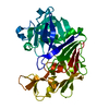
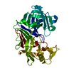
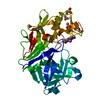
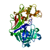
| Entry | Database: PDB / ID: 3pmy | ||||||
|---|---|---|---|---|---|---|---|
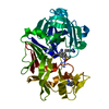
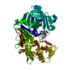
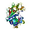
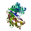
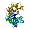
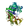
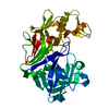
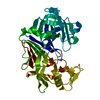
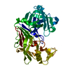
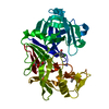
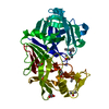
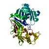
| Title | Endothiapepsin in complex with a fragment | ||||||
 Components Components | Endothiapepsin | ||||||
 Keywords Keywords | HYDROLASE/HYDROLASE INHIBITOR / HYDROLASE-HYDROLASE INHIBITOR complex | ||||||
| Function / homology |  Function and homology information Function and homology information | ||||||
| Biological species |  Cryphonectria parasitica (chestnut blight fungus) Cryphonectria parasitica (chestnut blight fungus) | ||||||
| Method |  X-RAY DIFFRACTION / SYNCHROTRON / MOLECULAR REPLACEMENT / Resolution: 1.38 Å X-RAY DIFFRACTION / SYNCHROTRON / MOLECULAR REPLACEMENT / Resolution: 1.38 Å | ||||||
 Authors Authors | Koester, H. / Heine, A. / Klebe, G. | ||||||
 Citation Citation | Journal: J.Med.Chem. / Year: 2011 Title: A small nonrule of 3 compatible fragment library provides high hit rate of endothiapepsin crystal structures with various fragment chemotypes. Authors: Koster, H. / Craan, T. / Brass, S. / Herhaus, C. / Zentgraf, M. / Neumann, L. / Heine, A. / Klebe, G. | ||||||
| History |
|
- Structure visualization
| Structure viewer | Molecule: MolmilJmol/JSmol |
|---|
- Downloads & links
-Download
| PDBx/mmCIF format | 3pmy.cif.gz | 142.3 KB | Display | PDBx/mmCIF format |
|---|---|---|---|---|
| PDB format | pdb3pmy.ent.gz | 110.5 KB | Display | PDB format |
| PDBx/mmJSON format | 3pmy.json.gz | Tree view | PDBx/mmJSON format | |
| Others |  Other downloads Other downloads |
-Validation report
| Summary document | 3pmy_validation.pdf.gz | 724.9 KB | Display | wwPDB validaton report |
|---|---|---|---|---|
| Full document | 3pmy_full_validation.pdf.gz | 726 KB | Display | |
| Data in XML | 3pmy_validation.xml.gz | 16.2 KB | Display | |
| Data in CIF | 3pmy_validation.cif.gz | 24.7 KB | Display | |
| Arichive directory | https://data.pdbj.org/pub/pdb/validation_reports/pm/3pmyftp://data.pdbj.org/pub/pdb/validation_reports/pm/3pmy | HTTPS FTP |
-Related structure data
| Related structure data |  3pb5C  3pbdC  3pbzC 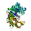 3pcwC  3pgiC 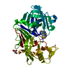 3pi0C 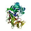 3pldC  3pllC 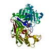 3pm4C  3pmuC  1oewS C: citing same article ( S: Starting model for refinement |
|---|---|
| Similar structure data |


-Links
 PDBj
PDBj
- Assembly
| Deposited unit | 
| ||||||||
|---|---|---|---|---|---|---|---|---|---|
| 1 |
| ||||||||
| Unit cell |
|
-Components
| #1: Protein | Mass: 33813.855 Da / Num. of mol.: 1 / Source method: isolated from a natural source Source: (natural) Cryphonectria parasitica (chestnut blight fungus)References: UniProt: P11838, endothiapepsin |
|---|---|
| #2: Chemical | ChemComp-41L /   Mass: 251.283 Da / Num. of mol.: 1 / Source method: obtained synthetically / Formula: C15H13N3O Mass: 251.283 Da / Num. of mol.: 1 / Source method: obtained synthetically / Formula: C15H13N3O |
| #3: Chemical | ChemComp-GOL /   Mass: 92.094 Da / Num. of mol.: 1 / Source method: obtained synthetically / Formula: C3H8O3 Mass: 92.094 Da / Num. of mol.: 1 / Source method: obtained synthetically / Formula: C3H8O3 |
| #4: Water | ChemComp-HOH /  Mass: 18.015 Da / Num. of mol.: 292 / Source method: isolated from a natural source / Formula: H2O Mass: 18.015 Da / Num. of mol.: 292 / Source method: isolated from a natural source / Formula: H2O |
| Has protein modification | Y |
-Experimental details
-Experiment
| Experiment | Method: X-RAY DIFFRACTION / Number of used crystals: 1 |
|---|
- Sample preparation
| Crystal | Density Matthews: 2.44 Å3/Da / Density % sol: 49.6 % |
|---|---|
| Crystal grow | Temperature: 289 K / Method: vapor diffusion, sitting drop / pH: 4.6 Details: 0.1M NH4Ac, 0.1M Acetate Buffer pH 4.6, 26% PEG 4000, VAPOR DIFFUSION, SITTING DROP, temperature 277K, temperature 289K |
-Data collection
| Diffraction | Mean temperature: 100 K |
|---|---|
| Diffraction source | Source: SYNCHROTRON / Site: BESSY  / Beamline: 14.2 / Wavelength: 0.91841 Å / Beamline: 14.2 / Wavelength: 0.91841 Å |
| Detector | Type: RAYONIX MX-225 / Detector: CCD / Date: Oct 21, 2010 / Details: mirrors |
| Radiation | Monochromator: Double Crystal Monochromator KMC-2 / Protocol: SINGLE WAVELENGTH / Monochromatic (M) / Laue (L): M / Scattering type: x-ray |
| Radiation wavelength | Wavelength: 0.91841 Å / Relative weight: 1 |
| Reflection | Resolution: 1.38→30 Å / Num. all: 65186 / Num. obs: 65186 / % possible obs: 100 % / Redundancy: 3 % / Rsym value: 0.046 / Net I/σ(I): 22.1 |
| Reflection shell | Resolution: 1.38→1.4 Å / Redundancy: 2 % / Mean I/σ(I) obs: 3.8 / Num. unique all: 2882 / Rsym value: 0.219 / % possible all: 100 |
- Processing
| Software |
| |||||||||||||||||||||||||||||||||
|---|---|---|---|---|---|---|---|---|---|---|---|---|---|---|---|---|---|---|---|---|---|---|---|---|---|---|---|---|---|---|---|---|---|---|
| Refinement | Method to determine structure: MOLECULAR REPLACEMENT Starting model: pdb entry 1OEW Resolution: 1.38→10 Å / Num. parameters: 24384 / Num. restraintsaints: 30397 / Cross valid method: FREE R / σ(F): 0 / Stereochemistry target values: Engh & Huber
| |||||||||||||||||||||||||||||||||
| Refine analyze | Num. disordered residues: 6 / Occupancy sum hydrogen: 2240 / Occupancy sum non hydrogen: 2686 | |||||||||||||||||||||||||||||||||
| Refinement step | Cycle: LAST / Resolution: 1.38→10 Å
| |||||||||||||||||||||||||||||||||
| Refine LS restraints |
|
