 Movie
Movie Controller

[English] 日本語
 Yorodumi
- PDB-3pi1: Crystallographic Structure of HbII-oxy from Lucina pectinata at pH 9.0 -
+ Open data

- Basic information
| Entry | Database: PDB / ID: 3pi1 | ||||||
|---|---|---|---|---|---|---|---|
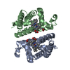
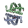
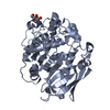
| Title | Crystallographic Structure of HbII-oxy from Lucina pectinata at pH 9.0 | ||||||
 Components Components | Hemoglobin II | ||||||
 Keywords Keywords | OXYGEN TRANSPORT / pH behavior / Oxygen Carrier | ||||||
| Function / homology |  Function and homology information Function and homology informationoxygen carrier activity / oxygen binding / heme binding / metal ion binding / cytoplasm Similarity search - Function | ||||||
| Biological species |  Lucina pectinata (invertebrata) Lucina pectinata (invertebrata) | ||||||
| Method |  X-RAY DIFFRACTION / SYNCHROTRON / MOLECULAR REPLACEMENT / molecular replacement / Resolution: 2.002 Å X-RAY DIFFRACTION / SYNCHROTRON / MOLECULAR REPLACEMENT / molecular replacement / Resolution: 2.002 Å | ||||||
 Authors Authors | Gavira, J.A. / Nieves-Marrero, C.A. / Ruiz-Martinez, C.R. / Estremera-Andujar, R.A. / Lopez-Garriga, J. / Garcia-Ruiz, J.M. | ||||||
 Citation Citation | Journal: To be Published Title: pH-dependence crystallographic studies of the oxygen carrier hemoglobin II from Lucina pectinata Authors: Nieves-Marrero, C.A. / Ruiz-Martinez, C.R. / Estremera-Andujar, R.A. / Lopez-Garriga, J. / Garcia-Ruiz, J.M. / Gavira, J.A. #1: Journal: Acta Crystallogr.,Sect.F / Year: 2010Title: Two-step counterdiffusion protocol for the crystallization of heamoglobin II from Lucina pectinata in the pH range 4-9 Authors: Nieves-Marrero, C.A. / Ruiz-Martinez, C.R. / Estremera-Andujar, R.A. / Gonzalez-Ramirez, L.A. / Lopez-Garriga, J. / Gavira, J.A. | ||||||
| History |
|
- Structure visualization
| Structure viewer | Molecule: MolmilJmol/JSmol |
|---|
- Downloads & links
-Download
| PDBx/mmCIF format | 3pi1.cif.gz | 81.9 KB | Display | PDBx/mmCIF format |
|---|---|---|---|---|
| PDB format | pdb3pi1.ent.gz | 61.4 KB | Display | PDB format |
| PDBx/mmJSON format | 3pi1.json.gz | Tree view | PDBx/mmJSON format | |
| Others |  Other downloads Other downloads |
-Validation report
| Summary document | 3pi1_validation.pdf.gz | 1.1 MB | Display | wwPDB validaton report |
|---|---|---|---|---|
| Full document | 3pi1_full_validation.pdf.gz | 1.1 MB | Display | |
| Data in XML | 3pi1_validation.xml.gz | 18.7 KB | Display | |
| Data in CIF | 3pi1_validation.cif.gz | 25.1 KB | Display | |
| Arichive directory | https://data.pdbj.org/pub/pdb/validation_reports/pi/3pi1ftp://data.pdbj.org/pub/pdb/validation_reports/pi/3pi1 | HTTPS FTP |
-Related structure data


-Links
 PDBj
PDBj


- Assembly
| Deposited unit | 
| ||||||||
|---|---|---|---|---|---|---|---|---|---|
| 1 |
| ||||||||
| 2 | 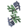
| ||||||||
| 3 | 
| ||||||||
| Unit cell |
|
-Components
| #1: Protein | Mass: 17041.557 Da / Num. of mol.: 2 / Source method: isolated from a natural source / Source: (natural) Lucina pectinata (invertebrata) / References: UniProt: Q86G74, UniProt: P41261*PLUS#2: Chemical |   Mass: 616.487 Da / Num. of mol.: 2 / Source method: obtained synthetically / Formula: C34H32FeN4O4 Mass: 616.487 Da / Num. of mol.: 2 / Source method: obtained synthetically / Formula: C34H32FeN4O4#3: Chemical |   Mass: 31.999 Da / Num. of mol.: 2 / Source method: obtained synthetically / Formula: O2 Mass: 31.999 Da / Num. of mol.: 2 / Source method: obtained synthetically / Formula: O2#4: Water | ChemComp-HOH / |  Mass: 18.015 Da / Num. of mol.: 224 / Source method: isolated from a natural source / Formula: H2O Mass: 18.015 Da / Num. of mol.: 224 / Source method: isolated from a natural source / Formula: H2OHas protein modification | Y | |
|---|
-Experimental details
-Experiment
| Experiment | Method: X-RAY DIFFRACTION / Number of used crystals: 1 |
|---|
- Sample preparation
| Crystal | Density Matthews: 3.24 Å3/Da / Density % sol: 62.01 % |
|---|---|
| Crystal grow | Temperature: 293 K / Method: capillary counterdiffusion / pH: 9 Details: Sodium Formate, pH 9.0, Capillary Counterdiffusion, temperature 293.0K |
-Data collection
| Diffraction | Mean temperature: 100 K |
|---|---|
| Diffraction source | Source: SYNCHROTRON / Site: ESRF  / Beamline: BM16 / Wavelength: 0.9075 Å / Beamline: BM16 / Wavelength: 0.9075 Å |
| Detector | Type: ADSC QUANTUM 4r / Detector: CCD / Date: Nov 2, 2008 |
| Radiation | Protocol: SINGLE WAVELENGTH / Monochromatic (M) / Laue (L): M / Scattering type: x-ray |
| Radiation wavelength | Wavelength: 0.9075 Å / Relative weight: 1 |
| Reflection | Resolution: 2→20 Å / Num. all: 29432 / Num. obs: 29432 / % possible obs: 95.5 % / Observed criterion σ(F): 0 / Observed criterion σ(I): -3 / Redundancy: 4.3 % / Rmerge(I) obs: 0.13 / Rsym value: 0.13 / Net I/σ(I): 7.78 |
| Reflection shell | Resolution: 2→2.07 Å / Redundancy: 4 % / Rmerge(I) obs: 0.512 / Mean I/σ(I) obs: 1.943 / Rsym value: 0.512 / % possible all: 98.1 |
| Cell measurement | Reflection used: 126200 |
-Phasing
| Phasing | Method: molecular replacement | |||||||||
|---|---|---|---|---|---|---|---|---|---|---|
| Phasing MR |
|
- Processing
| Software |
| ||||||||||||||||||||||||||||||||||||||||||||||||||||||||||||||||||||||
|---|---|---|---|---|---|---|---|---|---|---|---|---|---|---|---|---|---|---|---|---|---|---|---|---|---|---|---|---|---|---|---|---|---|---|---|---|---|---|---|---|---|---|---|---|---|---|---|---|---|---|---|---|---|---|---|---|---|---|---|---|---|---|---|---|---|---|---|---|---|---|---|
| Refinement | Method to determine structure: MOLECULAR REPLACEMENT / Resolution: 2.002→19.509 Å / Cor.coef. Fo:Fc: 0.961 / Cor.coef. Fo:Fc free: 0.942 / Occupancy max: 1 / Occupancy min: 0.22 / SU ML: 0.27 / σ(F): 0.09 / Phase error: 23.96 / Stereochemistry target values: ML Details: HYDROGENS HAVE BEEN ADDED IN THE RIDING POSITIONS U VALUES : REFINED INDIVIDUALLY
| ||||||||||||||||||||||||||||||||||||||||||||||||||||||||||||||||||||||
| Solvent computation | Shrinkage radii: 0.9 Å / VDW probe radii: 1.11 Å / Solvent model: FLAT BULK SOLVENT MODEL / Bsol: 59.558 Å2 / ksol: 0.42 e/Å3 | ||||||||||||||||||||||||||||||||||||||||||||||||||||||||||||||||||||||
| Displacement parameters |
| ||||||||||||||||||||||||||||||||||||||||||||||||||||||||||||||||||||||
| Refinement step | Cycle: LAST / Resolution: 2.002→19.509 Å
| ||||||||||||||||||||||||||||||||||||||||||||||||||||||||||||||||||||||
| Refine LS restraints |
| ||||||||||||||||||||||||||||||||||||||||||||||||||||||||||||||||||||||
| LS refinement shell |
|
