 Movie
Movie Controller

[English] 日本語
 Yorodumi
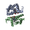
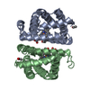
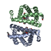
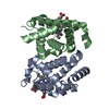
- PDB-6otw: Crystallographic Structure of (HbII-HbIII)-O2 from Lucina pectina... -
+ Open data

- Basic information
| Entry | Database: PDB / ID: 6otw | ||||||
|---|---|---|---|---|---|---|---|
| Title | Crystallographic Structure of (HbII-HbIII)-O2 from Lucina pectinata at pH 5.0 | ||||||
 Components Components |
| ||||||
 Keywords Keywords | OXYGEN TRANSPORT / hemeprotein | ||||||
| Function / homology |  Function and homology information Function and homology informationoxygen carrier activity / oxygen binding / heme binding / metal ion binding / cytoplasm Similarity search - Function | ||||||
| Biological species |  Phacoides pectinatus (invertebrata) Phacoides pectinatus (invertebrata) | ||||||
| Method |  X-RAY DIFFRACTION / SYNCHROTRON / MOLECULAR REPLACEMENT / Resolution: 2.447 Å X-RAY DIFFRACTION / SYNCHROTRON / MOLECULAR REPLACEMENT / Resolution: 2.447 Å | ||||||
 Authors Authors | Marchany-Rivera, D. / Smith, C.A. / Rodriguez-Perez, J.D. / Lopez-Garriga, J. | ||||||
| Funding support |  United States, 1items United States, 1items
| ||||||
 Citation Citation | Journal: J.Inorg.Biochem. / Year: 2020 Title: Lucina pectinata oxyhemoglobin (II-III) heterodimer pH susceptibility. Authors: Marchany-Rivera, D. / Smith, C.A. / Rodriguez-Perez, J.D. / Lopez-Garriga, J. | ||||||
| History |
|
- Structure visualization
| Structure viewer | Molecule: MolmilJmol/JSmol |
|---|
- Downloads & links
-Download
| PDBx/mmCIF format | 6otw.cif.gz | 143.8 KB | Display | PDBx/mmCIF format |
|---|---|---|---|---|
| PDB format | pdb6otw.ent.gz | 111.2 KB | Display | PDB format |
| PDBx/mmJSON format | 6otw.json.gz | Tree view | PDBx/mmJSON format | |
| Others |  Other downloads Other downloads |
-Validation report
| Arichive directory | https://data.pdbj.org/pub/pdb/validation_reports/ot/6otwftp://data.pdbj.org/pub/pdb/validation_reports/ot/6otw | HTTPS FTP |
|---|
-Related structure data
| Related structure data |  6otxC  6otyC  3pt8S S: Starting model for refinement C: citing same article ( |
|---|---|
| Similar structure data |


-Links
 PDBj
PDBj


- Assembly
| Deposited unit | 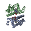
| ||||||||
|---|---|---|---|---|---|---|---|---|---|
| 1 |
| ||||||||
| Unit cell |
|
-Components
| #1: Protein | Mass: 17146.715 Da / Num. of mol.: 1 Source method: isolated from a genetically manipulated source Source: (gene. exp.) Phacoides pectinatus (invertebrata) / Production host: Phacoides pectinatus (invertebrata) / References: UniProt: Q86G74, UniProt: P41261*PLUS | ||
|---|---|---|---|
| #2: Protein | Mass: 17450.910 Da / Num. of mol.: 1 Source method: isolated from a genetically manipulated source Source: (gene. exp.) Phacoides pectinatus (invertebrata) / Production host: Phacoides pectinatus (invertebrata) / References: UniProt: P41262 | ||
| #3: Chemical |   Mass: 616.487 Da / Num. of mol.: 2 / Source method: obtained synthetically / Formula: C34H32FeN4O4 / Feature type: SUBJECT OF INVESTIGATION Mass: 616.487 Da / Num. of mol.: 2 / Source method: obtained synthetically / Formula: C34H32FeN4O4 / Feature type: SUBJECT OF INVESTIGATION#4: Water | ChemComp-HOH / |  Mass: 18.015 Da / Num. of mol.: 38 / Source method: isolated from a natural source / Formula: H2O Mass: 18.015 Da / Num. of mol.: 38 / Source method: isolated from a natural source / Formula: H2O |
-Experimental details
-Experiment
| Experiment | Method: X-RAY DIFFRACTION / Number of used crystals: 1 |
|---|
- Sample preparation
| Crystal | Density Matthews: 3.05 Å3/Da / Density % sol: 59.66 % |
|---|---|
| Crystal grow | Temperature: 293 K / Method: counter-diffusion / pH: 5 / Details: sodium formate |
-Data collection
| Diffraction | Mean temperature: 100 K / Serial crystal experiment: N |
|---|---|
| Diffraction source | Source: SYNCHROTRON / Site: SSRL / Beamline: BL9-2 / Wavelength: 1.19499 Å |
| Detector | Type: DECTRIS PILATUS 6M / Detector: PIXEL / Date: Jul 12, 2017 |
| Radiation | Protocol: SINGLE WAVELENGTH / Monochromatic (M) / Laue (L): M / Scattering type: x-ray |
| Radiation wavelength | Wavelength: 1.19499 Å / Relative weight: 1 |
| Reflection | Resolution: 2.447→50 Å / Num. obs: 16508 / % possible obs: 97.5 % / Redundancy: 6.9 % / Net I/σ(I): 26.7 |
| Reflection shell | Resolution: 2.447→2.49 Å / Num. unique obs: 262087 |
- Processing
| Software |
| |||||||||||||||||||||||||||||||||||||||||||||||||
|---|---|---|---|---|---|---|---|---|---|---|---|---|---|---|---|---|---|---|---|---|---|---|---|---|---|---|---|---|---|---|---|---|---|---|---|---|---|---|---|---|---|---|---|---|---|---|---|---|---|---|
| Refinement | Method to determine structure: MOLECULAR REPLACEMENT Starting model: 3PT8 Resolution: 2.447→33.228 Å / SU ML: 0.38 / Cross valid method: THROUGHOUT / σ(F): 1.34 / Phase error: 30.17
| |||||||||||||||||||||||||||||||||||||||||||||||||
| Solvent computation | Shrinkage radii: 0.9 Å / VDW probe radii: 1.11 Å | |||||||||||||||||||||||||||||||||||||||||||||||||
| Displacement parameters | Biso max: 167.83 Å2 / Biso mean: 75.2282 Å2 / Biso min: 36.77 Å2 | |||||||||||||||||||||||||||||||||||||||||||||||||
| Refinement step | Cycle: final / Resolution: 2.447→33.228 Å
| |||||||||||||||||||||||||||||||||||||||||||||||||
| LS refinement shell | Refine-ID: X-RAY DIFFRACTION / Rfactor Rfree error: 0 / Total num. of bins used: 6
| |||||||||||||||||||||||||||||||||||||||||||||||||
| Refinement TLS params. | Method: refined / Origin x: -9.6945 Å / Origin y: 12.9284 Å / Origin z: 24.5415 Å
| |||||||||||||||||||||||||||||||||||||||||||||||||
| Refinement TLS group |
|
