 Movie
Movie Controller

[English] 日本語
 Yorodumi
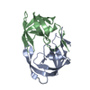
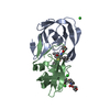
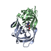
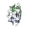
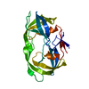
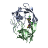
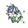
- PDB-3phv: X-RAY ANALYSIS OF HIV-1 PROTEINASE AT 2.7 ANGSTROMS RESOLUTION CO... -
+ Open data

- Basic information
| Entry | Database: PDB / ID: 3phv | ||||||
|---|---|---|---|---|---|---|---|
| Title | X-RAY ANALYSIS OF HIV-1 PROTEINASE AT 2.7 ANGSTROMS RESOLUTION CONFIRMS STRUCTURAL HOMOLOGY AMONG RETROVIRAL ENZYMES | ||||||
 Components Components | UNLIGANDED HIV-1 PROTEASE | ||||||
 Keywords Keywords | HYDROLASE / ASPARTIC PROTEINASE | ||||||
| Function / homology |  Function and homology information Function and homology informationintegrase activity / Integration of viral DNA into host genomic DNA / Autointegration results in viral DNA circles / Minus-strand DNA synthesis / Plus-strand DNA synthesis / Uncoating of the HIV Virion / 2-LTR circle formation / Vpr-mediated nuclear import of PICs / Early Phase of HIV Life Cycle / Integration of provirus ...integrase activity / Integration of viral DNA into host genomic DNA / Autointegration results in viral DNA circles / Minus-strand DNA synthesis / Plus-strand DNA synthesis / Uncoating of the HIV Virion / 2-LTR circle formation / Vpr-mediated nuclear import of PICs / Early Phase of HIV Life Cycle / Integration of provirus / APOBEC3G mediated resistance to HIV-1 infection / Binding and entry of HIV virion / viral life cycle / HIV-1 retropepsin / symbiont-mediated activation of host apoptosis / retroviral ribonuclease H / exoribonuclease H / exoribonuclease H activity / Assembly Of The HIV Virion / protein processing / viral genome integration into host DNA / Budding and maturation of HIV virion / establishment of integrated proviral latency / RNA-directed DNA polymerase / RNA stem-loop binding / viral penetration into host nucleus / host multivesicular body / RNA-directed DNA polymerase activity / RNA-DNA hybrid ribonuclease activity / Transferases; Transferring phosphorus-containing groups; Nucleotidyltransferases / peptidase activity / host cell / viral nucleocapsid / DNA recombination / DNA-directed DNA polymerase / aspartic-type endopeptidase activity / Hydrolases; Acting on ester bonds / DNA-directed DNA polymerase activity / symbiont-mediated suppression of host gene expression / viral translational frameshifting / symbiont entry into host cell / lipid binding / host cell nucleus / host cell plasma membrane / virion membrane / structural molecule activity / DNA binding / zinc ion binding / identical protein binding Similarity search - Function | ||||||
| Biological species |  HIV-1 M:B_HXB2R (virus) HIV-1 M:B_HXB2R (virus) | ||||||
| Method |  X-RAY DIFFRACTION / Resolution: 2.7 Å X-RAY DIFFRACTION / Resolution: 2.7 Å | ||||||
 Authors Authors | Lapatto, R. / Blundell, T.L. / Hemmings, A. / Wilderspin, A. / Wood, S.P. / Danley, D.E. / Geoghegan, K.F. / Hawrylik, S.J. / Hobart, P.M. | ||||||
 Citation Citation | Journal: Nature / Year: 1989 Title: X-ray analysis of HIV-1 proteinase at 2.7 A resolution confirms structural homology among retroviral enzymes. Authors: Lapatto, R. / Blundell, T. / Hemmings, A. / Overington, J. / Wilderspin, A. / Wood, S. / Merson, J.R. / Whittle, P.J. / Danley, D.E. / Geoghegan, K.F. / Hawrylik, S.J. / Lee, S.E. / Scheld, K.G. / Hobart, P.M. #1: Journal: J.Biol.Chem. / Year: 1989Title: Crystallization of the Aspartyl Protease from the Human Immunodeficiency Virus, HIV-1 Authors: Mckeever, B.M. / Navia, M.A. / Fitzgerald, P.M.D. / Springer, J.P. / Leu, C.-T. / Heimbach, J.C. / Herber, W.K. / Sigal, I.S. / Darke, P.L. | ||||||
| History |
| ||||||
| Remark 700 | SHEET THE DIMER INTERFACE IS COMPOSED OF INTERDIGITATED N- AND C-TERMINAL STRANDS FROM BOTH ...SHEET THE DIMER INTERFACE IS COMPOSED OF INTERDIGITATED N- AND C-TERMINAL STRANDS FROM BOTH SUBUNITS FORMING A FOUR-STRANDED ANTI-PARALLEL BETA-SHEET, S2. APPLICATION OF THE TWO-FOLD ROTATION TO RESIDUES 1 TO 5 AND 95 TO 99 GENERATES RESIDUES 1' TO 5' AND 95' TO 99' RESPECTIVELY. BECAUSE OF LIMITATIONS IMPOSED BY THE PROTEIN DATA BANK FORMAT IT IS NOT POSSIBLE TO PRESENT THIS SHEET ON SHEET RECORDS. INSTEAD THIS SHEET IS SPECIFIED IN THIS REMARK. STRANDS 1 AND 3 ARE FROM THE MOLECULE IN THIS ENTRY AND STRANDS 2 AND 4 ARE FROM THE SYMMETRY RELATED MOLECULE. 1 S2 4 PRO 1 LEU 5 0 2 S2 4 CYS 95' PHE 99'-1 3 S2 4 CYS 95 PHE 99 -1 4 S2 4 PRO 1' LEU 5'-1 |
- Structure visualization
| Structure viewer | Molecule: MolmilJmol/JSmol |
|---|
- Downloads & links
-Download
| PDBx/mmCIF format | 3phv.cif.gz | 28.5 KB | Display | PDBx/mmCIF format |
|---|---|---|---|---|
| PDB format | pdb3phv.ent.gz | 18.3 KB | Display | PDB format |
| PDBx/mmJSON format | 3phv.json.gz | Tree view | PDBx/mmJSON format | |
| Others |  Other downloads Other downloads |
-Validation report
| Arichive directory | https://data.pdbj.org/pub/pdb/validation_reports/ph/3phvftp://data.pdbj.org/pub/pdb/validation_reports/ph/3phv | HTTPS FTP |
|---|
-Related structure data
| Similar structure data |
|---|


-Links
 PDBj
PDBj


- Assembly
| Deposited unit | 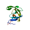
| ||||||||
|---|---|---|---|---|---|---|---|---|---|
| 1 | 
| ||||||||
| Unit cell |
|
-Components
| #1: Protein | Mass: 10803.756 Da / Num. of mol.: 1 Source method: isolated from a genetically manipulated source Source: (gene. exp.) HIV-1 M:B_HXB2R (virus) / Genus: Lentivirus / Species: Human immunodeficiency virus 1 / Cell line: S2 / Gene: POL / Organ: LEAVES / Gene (production host): POL / Production host:  |
|---|
-Experimental details
-Experiment
| Experiment | Method: X-RAY DIFFRACTION |
|---|
- Sample preparation
| Crystal | Density Matthews: 3.13 Å3/Da / Density % sol: 60.66 % | ||||||||||||||||||||||||||||||||||||||||||
|---|---|---|---|---|---|---|---|---|---|---|---|---|---|---|---|---|---|---|---|---|---|---|---|---|---|---|---|---|---|---|---|---|---|---|---|---|---|---|---|---|---|---|---|
| Crystal grow | *PLUS Temperature: 4 ℃ / pH: 7 / Method: vapor diffusion, hanging drop | ||||||||||||||||||||||||||||||||||||||||||
| Components of the solutions | *PLUS
|
-Data collection
| Radiation | Scattering type: x-ray |
|---|---|
| Radiation wavelength | Relative weight: 1 |
| Reflection | *PLUS Highest resolution: 2.7 Å / Num. obs: 2200 / Observed criterion σ(I): 2 / Num. measured all: 10000 / Rmerge(I) obs: 0.11 |
- Processing
| Software | Name: X-PLOR / Classification: refinement | ||||||||||||
|---|---|---|---|---|---|---|---|---|---|---|---|---|---|
| Refinement | Resolution: 2.7→10 Å / σ(I): 3 /
| ||||||||||||
| Refinement step | Cycle: LAST / Resolution: 2.7→10 Å
| ||||||||||||
| Refine LS restraints |
| ||||||||||||
| Refinement | *PLUS Highest resolution: 2.7 Å / Lowest resolution: 10 Å / Num. reflection obs: 2370 / σ(I): 3 / Rfactor obs: 0.191 | ||||||||||||
| Solvent computation | *PLUS | ||||||||||||
| Displacement parameters | *PLUS |
