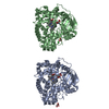
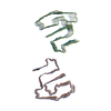
[English] 日本語
Yorodumi
- PDB-3pfq: Crystal Structure and Allosteric Activation of Protein Kinase C b... -

+
Open data


ID or keywords:

Loading...

-
Basic information

Entry
Database: PDB / ID: 3pfq
TitleCrystal Structure and Allosteric Activation of Protein Kinase C beta II
ComponentsProtein kinase C beta type
KeywordsTRANSFERASE / Kinase / Phosphorylation
Function / homology
Function and homology information


dibenzo-p-dioxin metabolic process / Depolymerization of the Nuclear Lamina / Disinhibition of SNARE formation / Response to elevated platelet cytosolic Ca2+ / Activation of NF-kappaB in B cells / histone H3T6 kinase activity / VEGFR2 mediated cell proliferation / positive regulation of odontogenesis of dentin-containing tooth / positive regulation of B cell receptor signaling pathway / spectrin ...dibenzo-p-dioxin metabolic process / Depolymerization of the Nuclear Lamina / Disinhibition of SNARE formation / Response to elevated platelet cytosolic Ca2+ / Activation of NF-kappaB in B cells / histone H3T6 kinase activity / VEGFR2 mediated cell proliferation / positive regulation of odontogenesis of dentin-containing tooth / positive regulation of B cell receptor signaling pathway / spectrin / regulation of glucose transmembrane transport / WNT5A-dependent internalization of FZD4 / RHO GTPases Activate NADPH Oxidases / protein kinase C / phospholipase C-activating G protein-coupled acetylcholine receptor signaling pathway / negative regulation of glucose transmembrane transport / cellular response to carbohydrate stimulus / diacylglycerol-dependent serine/threonine kinase activity / response to vitamin D / presynaptic cytosol / positive regulation of vascular endothelial growth factor receptor signaling pathway / regulation of growth / nuclear androgen receptor binding / regulation of synaptic vesicle exocytosis / B cell activation / regulation of dopamine secretion / Trafficking of GluR2-containing AMPA receptors / calyx of Held / calcium channel regulator activity / response to glucose / presynaptic modulation of chemical synaptic transmission / negative regulation of insulin receptor signaling pathway / post-translational protein modification / nuclear receptor coactivator activity / protein kinase C binding / brush border membrane / B cell receptor signaling pathway / positive regulation of insulin secretion / intracellular calcium ion homeostasis / positive regulation of angiogenesis / calcium ion transport / presynapse / histone binding / positive regulation of canonical NF-kappaB signal transduction / response to ethanol / adaptive immune response / intracellular signal transduction / response to xenobiotic stimulus / phosphorylation / protein serine kinase activity / protein serine/threonine kinase activity / centrosome / apoptotic process / chromatin binding / regulation of transcription by RNA polymerase II / zinc ion binding / nucleoplasm / ATP binding / membrane / nucleus / plasma membrane / cytoplasm
Similarity search - Function
Classical protein kinase C beta, catalytic domain / Protein kinase C, alpha/beta/gamma types / Protein kinase, C-terminal / Protein kinase C terminal domain / Diacylglycerol/phorbol-ester binding / C2 domain / Phorbol esters/diacylglycerol binding domain (C1 domain) / Zinc finger phorbol-ester/DAG-type signature. / C2 domain / Protein kinase C conserved region 2 (CalB) ...Classical protein kinase C beta, catalytic domain / Protein kinase C, alpha/beta/gamma types / Protein kinase, C-terminal / Protein kinase C terminal domain / Diacylglycerol/phorbol-ester binding / C2 domain / Phorbol esters/diacylglycerol binding domain (C1 domain) / Zinc finger phorbol-ester/DAG-type signature. / C2 domain / Protein kinase C conserved region 2 (CalB) / Zinc finger phorbol-ester/DAG-type profile. / Protein kinase C conserved region 1 (C1) domains (Cysteine-rich domains) / Protein kinase C-like, phorbol ester/diacylglycerol-binding domain / C2 domain / C2 domain profile. / C1-like domain superfamily / Extension to Ser/Thr-type protein kinases / AGC-kinase, C-terminal / AGC-kinase C-terminal domain profile. / C2 domain superfamily / Transferase(Phosphotransferase) domain 1 / Transferase(Phosphotransferase); domain 1 / Phosphorylase Kinase; domain 1 / Phosphorylase Kinase; domain 1 / Serine/threonine-protein kinase, active site / Serine/Threonine protein kinases active-site signature. / Protein kinase domain / Serine/Threonine protein kinases, catalytic domain / Protein kinase, ATP binding site / Protein kinases ATP-binding region signature. / Protein kinase domain profile. / Protein kinase domain / Protein kinase-like domain superfamily / Immunoglobulin-like / Sandwich / 2-Layer Sandwich / Orthogonal Bundle / Mainly Beta / Mainly Alpha / Alpha Beta
Similarity search - Domain/homology
PHOSPHOAMINOPHOSPHONIC ACID-ADENYLATE ESTER / Protein kinase C beta type
Similarity search - Component
Biological speciesRattus norvegicus (Norway rat)
MethodX-RAY DIFFRACTION / SYNCHROTRON / MOLECULAR REPLACEMENT / Resolution: 4 Å
AuthorsLeonard, T.A. / Rozycki, B. / Saidi, L.F. / Hummer, G. / Hurley, J.H.
CitationJournal: Cell(Cambridge,Mass.) / Year: 2011
Title: Crystal Structure and Allosteric Activation of Protein Kinase C beta II
Authors: Leonard, T.A. / Rozycki, B. / Saidi, L.F. / Hummer, G. / Hurley, J.H.
History
DepositionOct 28, 2010Deposition site: RCSB / Processing site: RCSB
Revision 1.0Feb 2, 2011Provider: repository / Type: Initial release
Revision 1.1Jul 13, 2011Group: Version format compliance
Revision 1.2Nov 8, 2017Group: Refinement description / Category: software / Item: _software.name
Revision 1.3Apr 11, 2018Group: Data collection / Database references ...Data collection / Database references / Derived calculations / Source and taxonomy / Structure summary
Category: diffrn_radiation_wavelength / diffrn_source ...diffrn_radiation_wavelength / diffrn_source / entity / entity_name_com / entity_src_gen / pdbx_struct_mod_residue / struct_ref / struct_ref_seq / struct_ref_seq_dif
Item: _diffrn_radiation_wavelength.wavelength / _diffrn_source.pdbx_wavelength_list ..._diffrn_radiation_wavelength.wavelength / _diffrn_source.pdbx_wavelength_list / _entity.pdbx_mutation / _entity_name_com.name / _entity_src_gen.gene_src_common_name / _entity_src_gen.pdbx_beg_seq_num / _entity_src_gen.pdbx_end_seq_num / _entity_src_gen.pdbx_seq_type / _pdbx_struct_mod_residue.details / _struct_ref.pdbx_db_isoform / _struct_ref.pdbx_seq_one_letter_code / _struct_ref_seq.db_align_end / _struct_ref_seq.pdbx_auth_seq_align_end / _struct_ref_seq.seq_align_end
Revision 1.4Sep 6, 2023Group: Data collection / Database references ...Data collection / Database references / Derived calculations / Refinement description
Category: chem_comp_atom / chem_comp_bond ...chem_comp_atom / chem_comp_bond / database_2 / pdbx_initial_refinement_model / pdbx_struct_conn_angle / struct_conn / struct_site
Item: _database_2.pdbx_DOI / _database_2.pdbx_database_accession ..._database_2.pdbx_DOI / _database_2.pdbx_database_accession / _pdbx_struct_conn_angle.ptnr1_auth_comp_id / _pdbx_struct_conn_angle.ptnr1_auth_seq_id / _pdbx_struct_conn_angle.ptnr1_label_atom_id / _pdbx_struct_conn_angle.ptnr1_label_comp_id / _pdbx_struct_conn_angle.ptnr1_label_seq_id / _pdbx_struct_conn_angle.ptnr3_auth_comp_id / _pdbx_struct_conn_angle.ptnr3_auth_seq_id / _pdbx_struct_conn_angle.ptnr3_label_atom_id / _pdbx_struct_conn_angle.ptnr3_label_comp_id / _pdbx_struct_conn_angle.ptnr3_label_seq_id / _pdbx_struct_conn_angle.value / _struct_conn.pdbx_dist_value / _struct_conn.pdbx_leaving_atom_flag / _struct_conn.ptnr1_auth_comp_id / _struct_conn.ptnr1_auth_seq_id / _struct_conn.ptnr1_label_atom_id / _struct_conn.ptnr1_label_comp_id / _struct_conn.ptnr1_label_seq_id / _struct_conn.ptnr2_auth_seq_id / _struct_conn.ptnr2_label_asym_id / _struct_site.pdbx_auth_asym_id / _struct_site.pdbx_auth_comp_id / _struct_site.pdbx_auth_seq_id

-
Structure visualization

Structure viewerMolecule:
MolmilJmol/JSmol

Downloads & links

-
Assembly

Deposited unit
A: Protein kinase C beta type
hetero molecules


Theoretical massNumber of molelcules
Total (without water)78,0117
Polymers77,2541
Non-polymers7576
Water00
1


  • Idetical with deposited unit
  • defined by author&software
TypeNameSymmetry operationNumber
identity operation1_555x,y,z1
MethodPISA
Unit cell
Length a, b, c (Å)114.270, 114.270, 170.840
Angle α, β, γ (deg.)90.00, 90.00, 120.00
Int Tables number154
Space group name H-MP3221

-
Components

#1: Protein Protein kinase C beta type / PKC-beta


Mass: 77253.859 Da / Num. of mol.: 1 / Fragment: PKC beta II / Mutation: C70S C217S C622S
Source method: isolated from a genetically manipulated source
Source: (gene. exp.) Rattus norvegicus (Norway rat) / Gene: Prkcb, Pkcb, Prkcb1 / Production host: 
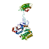
Trichoplusia ni (cabbage looper) / References: UniProt: P68403, protein kinase C
#2: Chemical ChemComp-CA / CALCIUM ION


Mass: 40.078 Da / Num. of mol.: 3 / Source method: obtained synthetically / Formula: Ca
#3: Chemical ChemComp-ZN / ZINC ION


Mass: 65.409 Da / Num. of mol.: 2 / Source method: obtained synthetically / Formula: Zn
#4: Chemical ChemComp-ANP / PHOSPHOAMINOPHOSPHONIC ACID-ADENYLATE ESTER


Mass: 506.196 Da / Num. of mol.: 1 / Source method: obtained synthetically / Formula: C10H17N6O12P3 / Comment: AMP-PNP, energy-carrying molecule analogue*YM
Sequence detailsTHE VARIANT SEQUENCE CORRESPONDS TO UNP P68403-2, ISOFORM BETA-II.

-
Experimental details

-
Experiment

ExperimentMethod: X-RAY DIFFRACTION / Number of used crystals: 1

-
Sample preparation

CrystalDensity Matthews: 4.17 Å3/Da / Density % sol: 70.49 %
Crystal growTemperature: 294 K / Method: vapor diffusion / pH: 8.5
Details: 0.1M Tris, pH 8.5, 3% PEG8K, VAPOR DIFFUSION, temperature 294K

-
Data collection

DiffractionMean temperature: 100 K
Diffraction sourceSource: SYNCHROTRON / Site: APS / Beamline: 23-ID-B / Wavelength: 1.03318 Å
DetectorType: MARMOSAIC 300 mm CCD / Detector: CCD / Date: Dec 4, 2009 / Details: mirrors
RadiationMonochromator: K-B pair of biomorph mirrors / Protocol: SINGLE WAVELENGTH / Monochromatic (M) / Laue (L): M / Scattering type: x-ray
Radiation wavelengthWavelength: 1.03318 Å / Relative weight: 1
ReflectionResolution: 4→57.1 Å / Num. all: 10670 / Num. obs: 10670 / % possible obs: 93.9 % / Observed criterion σ(F): 1.8 / Observed criterion σ(I): 1.8
Reflection shellResolution: 4→4.22 Å / % possible all: 88

-
Processing

Software
NameVersionClassification
JBluIce-EPICSdata collection
PHASERphasing
CNS1.3refinement
MOSFLMdata reduction
SCALAdata scaling
RefinementMethod to determine structure: MOLECULAR REPLACEMENT
Starting model: PDB entries 2I0E and 1A25
Resolution: 4→57.1 Å / σ(F): 1.8 / Stereochemistry target values: Engh & Huber
RfactorNum. reflectionSelection details
Rfree0.2445 508 RANDOM
Rwork0.1931 --
obs0.2445 10670 -
all-10670 -
Refinement stepCycle: LAST / Resolution: 4→57.1 Å
ProteinNucleic acidLigandSolventTotal
Num. atoms4237 0 36 0 4273
Refine LS restraints
Refine-IDTypeDev ideal
X-RAY DIFFRACTIONc_bond_d0.012
X-RAY DIFFRACTIONc_angle_deg0.878
LS refinement shellResolution: 4→4.14 Å
RfactorNum. reflection% reflection
Rfree0.2909 48 -
Rwork0.2673 --
obs-919 86.3 %

+
About Yorodumi

-
News

-
Feb 9, 2022. New format data for meta-information of EMDB entries

New format data for meta-information of EMDB entries

  • Version 3 of the EMDB header file is now the official format.
  • The previous official version 1.9 will be removed from the archive.

Related info.:EMDB header

External links:wwPDB to switch to version 3 of the EMDB data model

-
Aug 12, 2020. Covid-19 info

Covid-19 info

URL: https://pdbjlvh1.pdbj.org/emnavi/covid19.php

New page: Covid-19 featured information page in EM Navigator.

Related info.:Covid-19 info / Mar 5, 2020. Novel coronavirus structure data

+
Mar 5, 2020. Novel coronavirus structure data

Novel coronavirus structure data

Related info.:Yorodumi Speices / Aug 12, 2020. Covid-19 info

External links:COVID-19 featured content - PDBj / Molecule of the Month (242):Coronavirus Proteases

+
Jan 31, 2019. EMDB accession codes are about to change! (news from PDBe EMDB page)

EMDB accession codes are about to change! (news from PDBe EMDB page)

  • The allocation of 4 digits for EMDB accession codes will soon come to an end. Whilst these codes will remain in use, new EMDB accession codes will include an additional digit and will expand incrementally as the available range of codes is exhausted. The current 4-digit format prefixed with “EMD-” (i.e. EMD-XXXX) will advance to a 5-digit format (i.e. EMD-XXXXX), and so on. It is currently estimated that the 4-digit codes will be depleted around Spring 2019, at which point the 5-digit format will come into force.
  • The EM Navigator/Yorodumi systems omit the EMD- prefix.

Related info.:Q: What is EMD? / ID/Accession-code notation in Yorodumi/EM Navigator

External links:EMDB Accession Codes are Changing Soon! / Contact to PDBj

+
Jul 12, 2017. Major update of PDB

Major update of PDB

  • wwPDB released updated PDB data conforming to the new PDBx/mmCIF dictionary.
  • This is a major update changing the version number from 4 to 5, and with Remediation, in which all the entries are updated.
  • In this update, many items about electron microscopy experimental information are reorganized (e.g. em_software).
  • Now, EM Navigator and Yorodumi are based on the updated data.

External links:wwPDB Remediation / Enriched Model Files Conforming to OneDep Data Standards Now Available in the PDB FTP Archive

-
Yorodumi

Thousand views of thousand structures

  • Yorodumi is a browser for structure data from EMDB, PDB, SASBDB, etc.
  • This page is also the successor to EM Navigator detail page, and also detail information page/front-end page for Omokage search.
  • The word "yorodu" (or yorozu) is an old Japanese word meaning "ten thousand". "mi" (miru) is to see.

Related info.:EMDB / PDB / SASBDB / Comparison of 3 databanks / Yorodumi Search / Aug 31, 2016. New EM Navigator & Yorodumi / Yorodumi Papers / Jmol/JSmol / Function and homology information / Changes in new EM Navigator and Yorodumi

Read more