[English] 日本語
Yorodumi
- PDB-3p0a: X-ray structure of pentameric HIV-1 CA -

+
Open data


ID or keywords:

Loading...

-
Basic information

Entry
Database: PDB / ID: 3p0a
TitleX-ray structure of pentameric HIV-1 CA
ComponentsHIV-1 CA
KeywordsVIRAL PROTEIN / capsid protein
Function / homology
Function and homology information


viral budding via host ESCRT complex / HIV-1 retropepsin / symbiont-mediated activation of host apoptosis / retroviral ribonuclease H / exoribonuclease H / exoribonuclease H activity / host multivesicular body / DNA integration / viral genome integration into host DNA / RNA-directed DNA polymerase ...viral budding via host ESCRT complex / HIV-1 retropepsin / symbiont-mediated activation of host apoptosis / retroviral ribonuclease H / exoribonuclease H / exoribonuclease H activity / host multivesicular body / DNA integration / viral genome integration into host DNA / RNA-directed DNA polymerase / establishment of integrated proviral latency / telomerase activity / viral penetration into host nucleus / RNA stem-loop binding / RNA-DNA hybrid ribonuclease activity / Transferases; Transferring phosphorus-containing groups; Nucleotidyltransferases / host cell / viral nucleocapsid / DNA recombination / DNA-directed DNA polymerase / aspartic-type endopeptidase activity / Hydrolases; Acting on ester bonds / host cell cytoplasm / DNA-directed DNA polymerase activity / symbiont-mediated suppression of host gene expression / symbiont entry into host cell / viral translational frameshifting / lipid binding / host cell nucleus / host cell plasma membrane / virion membrane / structural molecule activity / proteolysis / DNA binding / RNA binding / zinc ion binding / membrane
Similarity search - Function
Gag protein p6 / Gag protein p6 / : / gag protein p24 N-terminal domain / Reverse transcriptase connection / Reverse transcriptase connection domain / Reverse transcriptase thumb / Reverse transcriptase thumb domain / Integrase Zinc binding domain / Zinc finger integrase-type profile. ...Gag protein p6 / Gag protein p6 / : / gag protein p24 N-terminal domain / Reverse transcriptase connection / Reverse transcriptase connection domain / Reverse transcriptase thumb / Reverse transcriptase thumb domain / Integrase Zinc binding domain / Zinc finger integrase-type profile. / Integrase-like, N-terminal / Integrase DNA binding domain / Integrase, C-terminal domain superfamily, retroviral / Integrase, N-terminal zinc-binding domain / Integrase, C-terminal, retroviral / Integrase DNA binding domain profile. / Immunodeficiency lentiviral matrix, N-terminal / gag gene protein p17 (matrix protein) / RNase H / Integrase core domain / Integrase, catalytic core / Integrase catalytic domain profile. / Retropepsin-like catalytic domain / Matrix protein, lentiviral and alpha-retroviral, N-terminal / Retroviral nucleocapsid Gag protein p24, C-terminal domain / Gag protein p24 C-terminal domain / RNase H type-1 domain profile. / Ribonuclease H domain / Retropepsins / Retroviral aspartyl protease / Aspartyl protease, retroviral-type family profile. / Peptidase A2A, retrovirus, catalytic / Reverse transcriptase domain / Reverse transcriptase (RNA-dependent DNA polymerase) / Reverse transcriptase (RT) catalytic domain profile. / Retrovirus capsid, C-terminal / Retroviral matrix protein / Retrovirus capsid, N-terminal / zinc finger / Zinc knuckle / Zinc finger, CCHC-type superfamily / Zinc finger, CCHC-type / Zinc finger CCHC-type profile. / Aspartic peptidase, active site / Eukaryotic and viral aspartyl proteases active site. / Aspartic peptidase domain superfamily / Ribonuclease H superfamily / Ribonuclease H-like superfamily / Reverse transcriptase/Diguanylate cyclase domain / DNA/RNA polymerase superfamily
Similarity search - Domain/homology
Gag-Pol polyprotein / Gag polyprotein
Similarity search - Component
Biological speciesHuman immunodeficiency virus 1
MethodX-RAY DIFFRACTION / SYNCHROTRON / MOLECULAR REPLACEMENT / Resolution: 5.954 Å
AuthorsPornillos, O.
CitationJournal: Nature / Year: 2011
Title: Atomic-level modelling of the HIV capsid.
Authors: Pornillos, O. / Ganser-Pornillos, B.K. / Yeager, M.
History
DepositionSep 27, 2010Deposition site: RCSB / Processing site: RCSB
Revision 1.0Jan 12, 2011Provider: repository / Type: Initial release
Revision 1.1Jul 13, 2011Group: Version format compliance
Revision 1.2Sep 6, 2023Group: Data collection / Database references / Refinement description
Category: chem_comp_atom / chem_comp_bond ...chem_comp_atom / chem_comp_bond / database_2 / pdbx_initial_refinement_model / struct_ref_seq_dif
Item: _database_2.pdbx_DOI / _database_2.pdbx_database_accession / _struct_ref_seq_dif.details

-
Structure visualization

Structure viewerMolecule:
MolmilJmol/JSmol

Downloads & links

-
Assembly

Deposited unit
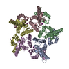
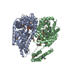
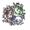
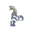
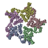
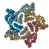
A: HIV-1 CA
B: HIV-1 CA
C: HIV-1 CA
D: HIV-1 CA
E: HIV-1 CA
F: HIV-1 CA
G: HIV-1 CA
H: HIV-1 CA
I: HIV-1 CA
J: HIV-1 CA
K: HIV-1 CA
L: HIV-1 CA
M: HIV-1 CA
N: HIV-1 CA
O: HIV-1 CA
P: HIV-1 CA
Q: HIV-1 CA
R: HIV-1 CA
S: HIV-1 CA
T: HIV-1 CA


Theoretical massNumber of molelcules
Total (without water)508,38420
Polymers508,38420
Non-polymers00
Water00
1
A: HIV-1 CA
B: HIV-1 CA
C: HIV-1 CA
D: HIV-1 CA
E: HIV-1 CA


Theoretical massNumber of molelcules
Total (without water)127,0965
Polymers127,0965
Non-polymers00
Water0
TypeNameSymmetry operationNumber
identity operation1_555x,y,z1
2
F: HIV-1 CA
G: HIV-1 CA
H: HIV-1 CA
I: HIV-1 CA
J: HIV-1 CA


Theoretical massNumber of molelcules
Total (without water)127,0965
Polymers127,0965
Non-polymers00
Water0
TypeNameSymmetry operationNumber
identity operation1_555x,y,z1
3
K: HIV-1 CA
L: HIV-1 CA
M: HIV-1 CA
N: HIV-1 CA
O: HIV-1 CA


Theoretical massNumber of molelcules
Total (without water)127,0965
Polymers127,0965
Non-polymers00
Water0
TypeNameSymmetry operationNumber
identity operation1_555x,y,z1
4
P: HIV-1 CA
Q: HIV-1 CA
R: HIV-1 CA
S: HIV-1 CA
T: HIV-1 CA


Theoretical massNumber of molelcules
Total (without water)127,0965
Polymers127,0965
Non-polymers00
Water0
TypeNameSymmetry operationNumber
identity operation1_555x,y,z1
Unit cell
Length a, b, c (Å)86.794, 122.128, 149.304
Angle α, β, γ (deg.)74.41, 74.36, 81.48
Int Tables number1
Space group name H-MP1

-
Components

#1: Protein
HIV-1 CA / Coordinate model: Cα atoms only


Mass: 25419.209 Da / Num. of mol.: 20 / Fragment: UNP residues 133-363 / Mutation: P17C, R18L, T19C, W184A, M185A
Source method: isolated from a genetically manipulated source
Source: (gene. exp.) Human immunodeficiency virus 1 / Strain: NL4-3 / Gene: CA, gag / Plasmid: pET11a / Production host: Escherichia coli (E. coli) / Strain (production host): BL21(DE3) / References: UniProt: Q72497, UniProt: P12497*PLUS
Compound detailsTHE REFINEMENT WAS LIMITED TO RIGID-BODY PLACEMENT OF THE MONOMERS. THE PROTEIN IS COMPOSED OF TWO ...THE REFINEMENT WAS LIMITED TO RIGID-BODY PLACEMENT OF THE MONOMERS. THE PROTEIN IS COMPOSED OF TWO DOMAINS, WHICH WERE TREATED AS SEPARATE RIGID BODIES DURING REFINEMENT. THE TWO DOMAINS WERE ALLOWED TO MOVE INDEPENDENTLY AND THAT IS WHY THE CONNECTIVITY BETWEEN THEM IS NOT WELL-PRESERVED.

-
Experimental details

-
Experiment

ExperimentMethod: X-RAY DIFFRACTION

-
Sample preparation

CrystalDensity Matthews: 2.88 Å3/Da / Density % sol: 57.26 %
Crystal growTemperature: 293 K / Method: vapor diffusion, sitting drop
Details: 30-32% PEG 2000 MME, 100 mM Tris, 0.4 M sodium iodide, pH 7-8, VAPOR DIFFUSION, SITTING DROP, temperature 293K
PH range: 7-8

-
Data collection

DiffractionMean temperature: 100 K
Diffraction sourceSource: SYNCHROTRON / Site: APS / Beamline: 22-ID / Wavelength: 1 Å
DetectorType: MARMOSAIC 300 mm CCD / Detector: CCD / Date: Mar 8, 2010
RadiationProtocol: SINGLE WAVELENGTH / Monochromatic (M) / Laue (L): M / Scattering type: x-ray
Radiation wavelengthWavelength: 1 Å / Relative weight: 1
ReflectionResolution: 5.95→50 Å / Num. all: 14299 / Num. obs: 14213 / % possible obs: 99.4 % / Observed criterion σ(F): 2 / Observed criterion σ(I): 2
Reflection shellResolution: 5.95→6.32 Å / Redundancy: 3.9 % / Mean I/σ(I) obs: 2.3 / Rsym value: 0.579 / % possible all: 98.6

-
Processing

Software
NameVersionClassification
MOLREPphasing
PHENIX(phenix.refine: 1.6.1_357)refinement
HKL-2000data reduction
HKL-2000data scaling
RefinementMethod to determine structure: MOLECULAR REPLACEMENT
Starting model: PDB entry 3P05
Resolution: 5.954→45.562 Å / SU ML: 1.08 / σ(F): 1.97 / Phase error: 36.41 / Stereochemistry target values: ML / Details: THE COORDINATES CONTAIN ONLY CALPHA ATOMS
RfactorNum. reflection% reflection
Rfree0.3156 1416 9.99 %
Rwork0.297 --
obs0.2989 14169 97.87 %
Solvent computationShrinkage radii: 0.9 Å / VDW probe radii: 1.11 Å / Solvent model: FLAT BULK SOLVENT MODEL / Bsol: 20 Å2 / ksol: 0.309 e/Å3
Displacement parameters
Baniso -1Baniso -2Baniso -3
1-279.7381 Å2-17.6802 Å217.3471 Å2
2--159.8556 Å2-9.2281 Å2
3---125.0104 Å2
Refinement stepCycle: LAST / Resolution: 5.954→45.562 Å
ProteinNucleic acidLigandSolventTotal
Num. atoms3952 0 0 0 3952
Refine LS restraints
Refine-IDTypeDev idealNumber
X-RAY DIFFRACTIONf_bond_d0.02930704
X-RAY DIFFRACTIONf_angle_d1.82741808
X-RAY DIFFRACTIONf_dihedral_angle_d15.56411256
X-RAY DIFFRACTIONf_chiral_restr0.0994856
X-RAY DIFFRACTIONf_plane_restr0.015368
LS refinement shell
Resolution (Å)Rfactor RfreeNum. reflection RfreeRfactor RworkNum. reflection RworkRefine-ID% reflection obs (%)
5.954-6.16670.38061420.39341120X-RAY DIFFRACTION86
6.1667-6.4130.41351420.39041265X-RAY DIFFRACTION99
6.413-6.7040.34671420.40071289X-RAY DIFFRACTION100
6.704-7.05620.36961420.36131311X-RAY DIFFRACTION100
7.0562-7.49650.33611420.32231303X-RAY DIFFRACTION100
7.4965-8.07240.30451420.31821306X-RAY DIFFRACTION100
8.0724-8.87930.27771420.28051307X-RAY DIFFRACTION100
8.8793-10.15180.2741420.23291287X-RAY DIFFRACTION100
10.1518-12.74360.26151410.23331292X-RAY DIFFRACTION98
12.7436-45.56370.35651390.32011273X-RAY DIFFRACTION98

+
About Yorodumi

-
News

-
Feb 9, 2022. New format data for meta-information of EMDB entries

New format data for meta-information of EMDB entries

  • Version 3 of the EMDB header file is now the official format.
  • The previous official version 1.9 will be removed from the archive.

Related info.:EMDB header

External links:wwPDB to switch to version 3 of the EMDB data model

-
Aug 12, 2020. Covid-19 info

Covid-19 info

URL: https://pdbj.org/emnavi/covid19.php

New page: Covid-19 featured information page in EM Navigator.

Related info.:Covid-19 info / Mar 5, 2020. Novel coronavirus structure data

+
Mar 5, 2020. Novel coronavirus structure data

Novel coronavirus structure data

Related info.:Yorodumi Speices / Aug 12, 2020. Covid-19 info

External links:COVID-19 featured content - PDBj / Molecule of the Month (242):Coronavirus Proteases

+
Jan 31, 2019. EMDB accession codes are about to change! (news from PDBe EMDB page)

EMDB accession codes are about to change! (news from PDBe EMDB page)

  • The allocation of 4 digits for EMDB accession codes will soon come to an end. Whilst these codes will remain in use, new EMDB accession codes will include an additional digit and will expand incrementally as the available range of codes is exhausted. The current 4-digit format prefixed with “EMD-” (i.e. EMD-XXXX) will advance to a 5-digit format (i.e. EMD-XXXXX), and so on. It is currently estimated that the 4-digit codes will be depleted around Spring 2019, at which point the 5-digit format will come into force.
  • The EM Navigator/Yorodumi systems omit the EMD- prefix.

Related info.:Q: What is EMD? / ID/Accession-code notation in Yorodumi/EM Navigator

External links:EMDB Accession Codes are Changing Soon! / Contact to PDBj

+
Jul 12, 2017. Major update of PDB

Major update of PDB

  • wwPDB released updated PDB data conforming to the new PDBx/mmCIF dictionary.
  • This is a major update changing the version number from 4 to 5, and with Remediation, in which all the entries are updated.
  • In this update, many items about electron microscopy experimental information are reorganized (e.g. em_software).
  • Now, EM Navigator and Yorodumi are based on the updated data.

External links:wwPDB Remediation / Enriched Model Files Conforming to OneDep Data Standards Now Available in the PDB FTP Archive

-
Yorodumi

Thousand views of thousand structures

  • Yorodumi is a browser for structure data from EMDB, PDB, SASBDB, etc.
  • This page is also the successor to EM Navigator detail page, and also detail information page/front-end page for Omokage search.
  • The word "yorodu" (or yorozu) is an old Japanese word meaning "ten thousand". "mi" (miru) is to see.

Related info.:EMDB / PDB / SASBDB / Comparison of 3 databanks / Yorodumi Search / Aug 31, 2016. New EM Navigator & Yorodumi / Yorodumi Papers / Jmol/JSmol / Function and homology information / Changes in new EM Navigator and Yorodumi

Read more