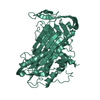
- PDB-3oz2: Crystal structure of a geranylgeranyl bacteriochlorophyll reducta... -
+
Open data
ID or keywords:
Loading...
-
Basic information
Entry
Database: PDB / ID: 3oz2
Title
Crystal structure of a geranylgeranyl bacteriochlorophyll reductase-like (Ta0516) from Thermoplasma acidophilum at 1.60 A resolution
Components
Digeranylgeranylglycerophospholipid reductase
Keywords
Flavoprotein / oxidoreductase / STRUCTURAL GENOMICS / JOINT CENTER FOR STRUCTURAL GENOMICS / JCSG / PROTEIN STRUCTURE INITIATIVE / PSI-BIOLOGY
Function / homology
Function and homology information
2,3-bis-O-geranylgeranyl-sn-glycerol 1-phosphate reductase [NAD(P)H] / : / glycerophospholipid biosynthetic process / : / oxidoreductase activity, acting on the CH-CH group of donors, NAD or NADP as acceptor / NAD binding / NADP binding / flavin adenine dinucleotide binding / plasma membrane Similarity search - Function
Mass: 44029.816 Da / Num. of mol.: 1 Source method: isolated from a genetically manipulated source Source: (gene. exp.) Thermoplasma acidophilum (acidophilic) / Gene: Ta0516 / Plasmid: SpeedET / Production host: Escherichia Coli (E. coli) / Strain (production host): HK100 References: UniProt: Q9HKS9, Oxidoreductases; Acting on the CH-CH group of donors; With NAD+ or NADP+ as acceptor
Mass: 18.015 Da / Num. of mol.: 332 / Source method: isolated from a natural source / Formula: H2O
-
Details
Has protein modification
Y
Sequence details
THE CONSTRUCT WAS EXPRESSED WITH A PURIFICATION TAG MGSDKIHHHHHHENLYFQG. THE TAG WAS REMOVED WITH ...THE CONSTRUCT WAS EXPRESSED WITH A PURIFICATION TAG MGSDKIHHHHHHENLYFQG. THE TAG WAS REMOVED WITH TEV PROTEASE LEAVING ONLY A GLYCINE FOLLOWED BY THE TARGET SEQUENCE.
-
Experimental details
-
Experiment
Experiment
Method: X-RAY DIFFRACTION / Number of used crystals: 1
-
Sample preparation
Crystal
Density Matthews: 2.35 Å3/Da / Density % sol: 47.55 %
Crystal grow
Temperature: 277 K / Method: vapor diffusion, sitting drop / pH: 7.5 Details: 15.00% Glycerol, 8.50% iso-Propanol, 17.00% PEG-4000, 0.1M HEPES pH 7.5, NANODROP, VAPOR DIFFUSION, SITTING DROP, temperature 277K
Resolution: 1.6→45.257 Å / Num. obs: 55071 / % possible obs: 99.4 % / Observed criterion σ(I): -3 / Biso Wilson estimate: 20.479 Å2 / Rmerge(I) obs: 0.052 / Net I/σ(I): 13.16
Reflection shell
Resolution (Å)
Rmerge(I) obs
Mean I/σ(I) obs
Num. measured obs
Num. unique obs
Diffraction-ID
% possible all
1.6-1.64
0.466
2.4
14404
4009
1
99.6
1.64-1.69
0.372
3
14078
3912
1
99.5
1.69-1.74
0.305
3.7
13773
3820
1
99.6
1.74-1.79
0.252
4.5
13485
3726
1
99.7
1.79-1.85
0.205
5.5
13195
3623
1
99.8
1.85-1.91
0.155
7.1
12708
3500
1
99.9
1.91-1.98
0.116
9.2
12328
3382
1
99.9
1.98-2.07
0.096
11.2
11996
3281
1
99.9
2.07-2.16
0.079
13.4
11420
3126
1
99.9
2.16-2.26
0.07
15.2
10887
2980
1
99.8
2.26-2.39
0.061
17.4
10483
2862
1
99.8
2.39-2.53
0.054
19.1
9911
2709
1
99.6
2.53-2.7
0.051
20.6
9339
2554
1
99.5
2.7-2.92
0.046
23.1
8633
2368
1
99.1
2.92-3.2
0.04
25.6
8021
2200
1
99.4
3.2-3.58
0.035
28.1
7215
1984
1
98.6
3.58-4.13
0.033
30.1
6360
1765
1
98.5
4.13-5.06
0.032
30.7
5337
1500
1
98
5.06-7.16
0.039
29.5
4104
1180
1
96.4
7.16-45.257
0.038
29.4
2120
648
1
89.4
-
Phasing
Phasing
Method: MAD
-
Processing
Software
Name
Version
Classification
NB
SHELX
phasing
REFMAC
5.5.0110
refinement
XSCALE
datascaling
PDB_EXTRACT
3.1
dataextraction
XDS
datareduction
SHELXD
phasing
autoSHARP
phasing
Refinement
Method to determine structure: MAD / Resolution: 1.6→45.257 Å / Cor.coef. Fo:Fc: 0.972 / Cor.coef. Fo:Fc free: 0.968 / Occupancy max: 1 / Occupancy min: 0.37 / SU B: 2.679 / SU ML: 0.046 / Cross valid method: THROUGHOUT / σ(F): 0 / ESU R: 0.079 / ESU R Free: 0.074 Stereochemistry target values: MAXIMUM LIKELIHOOD WITH PHASES Details: 1. HYDROGENS HAVE BEEN ADDED IN THE RIDING POSITIONS 2. A MET-INHIBITION PROTOCOL WAS USED FOR SELENOMETHIONINE INCORPORATION DURING PROTEIN EXPRESSION. THE OCCUPANCY OF THE SE ATOMS IN THE ...Details: 1. HYDROGENS HAVE BEEN ADDED IN THE RIDING POSITIONS 2. A MET-INHIBITION PROTOCOL WAS USED FOR SELENOMETHIONINE INCORPORATION DURING PROTEIN EXPRESSION. THE OCCUPANCY OF THE SE ATOMS IN THE MSE RESIDUES WAS REDUCED TO 0.75 FOR THE REDUCED SCATTERING POWER DUE TO PARTIAL S-MET INCORPORATION. 3. ATOM RECORD CONTAINS SUM OF TLS AND RESIDUAL B FACTORS. ANISOU RECORD CONTAINS SUM OF TLS AND RESIDUAL U FACTORS. 4. WATERS WERE EXCLUDED FROM AUTOMATIC TLS ASSIGNMENT. 5. FAD IS MODELED BASED ON DENSITY AND FUNCTION. 6. A BACTERIAL LIPID FOUND IN THE ACTIVE SITE WAS TENTATIVELY ASSIGNED AS A PHOSPHATIDYLGYLCEROL (OZ2) BASED ON DENSITY AND FUNCTION. THE DENSITY FOR THE HEAD GROUP AND LIPID TAILS ARE POORLY DEFINED. 7. ETHYLENE GLYCOL (EDO) AND GLYCEROL (GOL) WERE MODELED BASED ON CRYSTALLIZATION/CYRO CONDITIONS.
Rfactor
Num. reflection
% reflection
Selection details
Rfree
0.1706
2794
5.1 %
RANDOM
Rwork
0.153
-
-
-
obs
0.1539
55071
99.35 %
-
Solvent computation
Ion probe radii: 0.8 Å / Shrinkage radii: 0.8 Å / VDW probe radii: 1.2 Å / Solvent model: MASK
In the structure databanks used in Yorodumi, some data are registered as the other names, "COVID-19 virus" and "2019-nCoV". Here are the details of the virus and the list of structure data.
Jan 31, 2019. EMDB accession codes are about to change! (news from PDBe EMDB page)
EMDB accession codes are about to change! (news from PDBe EMDB page)
The allocation of 4 digits for EMDB accession codes will soon come to an end. Whilst these codes will remain in use, new EMDB accession codes will include an additional digit and will expand incrementally as the available range of codes is exhausted. The current 4-digit format prefixed with “EMD-” (i.e. EMD-XXXX) will advance to a 5-digit format (i.e. EMD-XXXXX), and so on. It is currently estimated that the 4-digit codes will be depleted around Spring 2019, at which point the 5-digit format will come into force.
The EM Navigator/Yorodumi systems omit the EMD- prefix.
Related info.:Q: What is EMD? / ID/Accession-code notation in Yorodumi/EM Navigator
Yorodumi is a browser for structure data from EMDB, PDB, SASBDB, etc.
This page is also the successor to EM Navigator detail page, and also detail information page/front-end page for Omokage search.
The word "yorodu" (or yorozu) is an old Japanese word meaning "ten thousand". "mi" (miru) is to see.
Related info.:EMDB / PDB / SASBDB / Comparison of 3 databanks / Yorodumi Search / Aug 31, 2016. New EM Navigator & Yorodumi / Yorodumi Papers / Jmol/JSmol / Function and homology information / Changes in new EM Navigator and Yorodumi
 Movie
Movie Controller

 Yorodumi
 Open data
 Basic information
 Components
 Keywords
 Function and homology information
 Thermoplasma acidophilum (acidophilic)
 X-RAY DIFFRACTION /
  Authors
 Citation
 Structure visualization
 Downloads & links
 Other downloads


 PDBj
PDBj Assembly


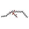


 Mass: 785.550 Da / Num. of mol.: 1 / Source method: obtained synthetically / Formula: C27H33N9O15P2 / Comment: FAD*YM
 Mass: 704.912 Da / Num. of mol.: 1 / Source method: obtained synthetically / Formula: C37H69O10P
 Mass: 62.068 Da / Num. of mol.: 11 / Source method: obtained synthetically / Formula: C2H6O2
 Mass: 92.094 Da / Num. of mol.: 2 / Source method: obtained synthetically / Formula: C3H8O3
 Sample preparation
 / Beamline: BL11-1 / Wavelength: 0.91837,0.97920,0.97862
 Processing