 Movie
Movie Controller

[English] 日本語
 Yorodumi
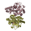
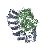
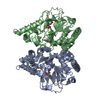
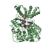
- PDB-3ox4: Structures of iron-dependent alcohol dehydrogenase 2 from Zymomon... -
+ Open data

- Basic information
| Entry | Database: PDB / ID: 3ox4 | ||||||
|---|---|---|---|---|---|---|---|
| Title | Structures of iron-dependent alcohol dehydrogenase 2 from Zymomonas mobilis ZM4 complexed with NAD cofactor | ||||||
 Components Components | Alcohol dehydrogenase 2 | ||||||
 Keywords Keywords | OXIDOREDUCTASE / Alcohol Dehydrogenase / Iron / NAD | ||||||
| Function / homology |  Function and homology information Function and homology informationethanol dehydrogenase (NAD+) activity / alcohol dehydrogenase (NAD+) activity / alcohol dehydrogenase / metal ion binding Similarity search - Function | ||||||
| Biological species |  Zymomonas mobilis (bacteria) Zymomonas mobilis (bacteria) | ||||||
| Method |  X-RAY DIFFRACTION / SYNCHROTRON / MOLECULAR REPLACEMENT / Resolution: 2 Å X-RAY DIFFRACTION / SYNCHROTRON / MOLECULAR REPLACEMENT / Resolution: 2 Å | ||||||
 Authors Authors | Moon, J.H. / Lee, H.J. / Song, J.M. / Park, S.Y. / Park, M.Y. / Park, H.M. / Sun, J. / Park, J.H. / Kim, J.S. | ||||||
 Citation Citation | Journal: J.Mol.Biol. / Year: 2011 Title: Structures of iron-dependent alcohol dehydrogenase 2 from Zymomonas mobilis ZM4 with and without NAD+ cofactor Authors: Moon, J.H. / Lee, H.J. / Park, S.Y. / Song, J.M. / Park, M.Y. / Park, H.M. / Sun, J. / Park, J.H. / Kim, B.Y. / Kim, J.S. | ||||||
| History |
|
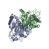
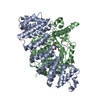
- Structure visualization
| Structure viewer | Molecule: MolmilJmol/JSmol |
|---|
- Downloads & links
-Download
| PDBx/mmCIF format | 3ox4.cif.gz | 298.2 KB | Display | PDBx/mmCIF format |
|---|---|---|---|---|
| PDB format | pdb3ox4.ent.gz | 242.5 KB | Display | PDB format |
| PDBx/mmJSON format | 3ox4.json.gz | Tree view | PDBx/mmJSON format | |
| Others |  Other downloads Other downloads |
-Validation report
| Arichive directory | https://data.pdbj.org/pub/pdb/validation_reports/ox/3ox4ftp://data.pdbj.org/pub/pdb/validation_reports/ox/3ox4 | HTTPS FTP |
|---|
-Related structure data
| Related structure data |  3owoSC S: Starting model for refinement C: citing same article ( |
|---|---|
| Similar structure data |


-Links
 PDBj
PDBj


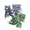
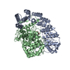
- Assembly
| Deposited unit | 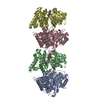
| ||||||||
|---|---|---|---|---|---|---|---|---|---|
| 1 | 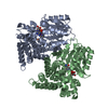
| ||||||||
| 2 | 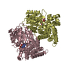
| ||||||||
| Unit cell |
|
-Components
| #1: Protein | Mass: 40186.098 Da / Num. of mol.: 4 Source method: isolated from a genetically manipulated source Source: (gene. exp.) Zymomonas mobilis (bacteria) / Strain: ZM4 / Gene: adhB, ZMO1596 / Production host: References: UniProt: P06758, UniProt: P0DJA2*PLUS, alcohol dehydrogenase #2: Chemical | ChemComp-NAD /   Mass: 663.425 Da / Num. of mol.: 4 / Source method: obtained synthetically / Formula: C21H27N7O14P2 / Comment: NAD*YM Mass: 663.425 Da / Num. of mol.: 4 / Source method: obtained synthetically / Formula: C21H27N7O14P2 / Comment: NAD*YM#3: Chemical | ChemComp-FE2 /   Mass: 55.845 Da / Num. of mol.: 4 / Source method: obtained synthetically / Formula: Fe Mass: 55.845 Da / Num. of mol.: 4 / Source method: obtained synthetically / Formula: Fe#4: Water | ChemComp-HOH / |  Mass: 18.015 Da / Num. of mol.: 555 / Source method: isolated from a natural source / Formula: H2O Mass: 18.015 Da / Num. of mol.: 555 / Source method: isolated from a natural source / Formula: H2O |
|---|
-Experimental details
-Experiment
| Experiment | Method: X-RAY DIFFRACTION / Number of used crystals: 1 |
|---|
- Sample preparation
| Crystal | Density Matthews: 2.03 Å3/Da / Density % sol: 39.42 % |
|---|---|
| Crystal grow | Temperature: 295 K / Method: vapor diffusion, hanging drop / pH: 8.5 Details: 25% PEG 4000, 0.15M Magnesium Chloride, 0.1M Tris-HCl, pH 8.5, VAPOR DIFFUSION, HANGING DROP, temperature 295.0K |
-Data collection
| Diffraction | Mean temperature: 100 K |
|---|---|
| Diffraction source | Source: SYNCHROTRON / Site: PAL/PLS  / Beamline: 4A / Wavelength: 1.2 Å / Beamline: 4A / Wavelength: 1.2 Å |
| Detector | Type: ADSC QUANTUM 315r / Detector: CCD / Date: Jun 22, 2010 |
| Radiation | Monochromator: GRAPHITE / Protocol: SINGLE WAVELENGTH / Monochromatic (M) / Laue (L): M / Scattering type: x-ray |
| Radiation wavelength | Wavelength: 1.2 Å / Relative weight: 1 |
| Reflection | Resolution: 2→50 Å / Num. obs: 84735 / % possible obs: 97.7 % / Observed criterion σ(F): -3 / Observed criterion σ(I): -3 / Redundancy: 2.7 % / Rsym value: 0.071 / Net I/σ(I): 15.3 |
| Reflection shell | Resolution: 2→2.03 Å / Redundancy: 2.4 % / Mean I/σ(I) obs: 2.37 / Num. unique all: 4206 / Rsym value: 0.404 / % possible all: 97 |
- Processing
| Software |
| |||||||||||||||||||||||||
|---|---|---|---|---|---|---|---|---|---|---|---|---|---|---|---|---|---|---|---|---|---|---|---|---|---|---|
| Refinement | Method to determine structure: MOLECULAR REPLACEMENT Starting model: PDB ENTRY 3OWO Resolution: 2→28.75 Å / Cross valid method: THROUGHOUT / σ(F): -3 / σ(I): -3 / Stereochemistry target values: Engh & Huber
| |||||||||||||||||||||||||
| Refinement step | Cycle: LAST / Resolution: 2→28.75 Å
| |||||||||||||||||||||||||
| Refine LS restraints |
|
