 Movie
Movie Controller

[English] 日本語
 Yorodumi
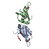
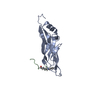
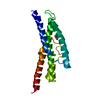
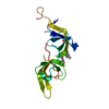
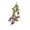
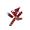
- PDB-3ov8: Crystal structure of AF1382 from Archaeoglobus fulgidus, High res... -
+ Open data

- Basic information
| Entry | Database: PDB / ID: 3ov8 | ||||||
|---|---|---|---|---|---|---|---|
| Title | Crystal structure of AF1382 from Archaeoglobus fulgidus, High resolution | ||||||
 Components Components | Protein AF_1382 | ||||||
 Keywords Keywords | UNKNOWN FUNCTION / AF1382 / PSI / STRUCTURAL GENOMICS / SOUTHEAST COLLABORATORY FOR STRUCTURAL GENOMICS / SECSG / putative DNS binding | ||||||
| Function / homology | Winged helix-like DNA-binding domain superfamily/Winged helix DNA-binding domain / Arc Repressor Mutant, subunit A / Winged helix DNA-binding domain superfamily / Winged helix-like DNA-binding domain superfamily / Orthogonal Bundle / Mainly Alpha / ACETATE ION / Uncharacterized protein AF_1382 Function and homology information Function and homology information | ||||||
| Biological species |   Archaeoglobus fulgidus (archaea) Archaeoglobus fulgidus (archaea) | ||||||
| Method |  X-RAY DIFFRACTION / SYNCHROTRON / FOURIER SYNTHESIS / Resolution: 1.8501 Å X-RAY DIFFRACTION / SYNCHROTRON / FOURIER SYNTHESIS / Resolution: 1.8501 Å | ||||||
 Authors Authors | Zhu, J.-Y. / Zhao, M. / Fu, Z.-Q. / Yang, H. / Chang, J. / Hao, X. / Chen, L. / Rose, J.P. / Wang, B.C. / Southeast Collaboratory for Structural Genomics (SECSG) | ||||||
 Citation Citation | Journal: Acta Crystallogr.,Sect.D / Year: 2012 Title: Structure of the Archaeoglobus fulgidus orphan ORF AF1382 determined by sulfur SAD from a moderately diffracting crystal. Authors: Zhu, J.Y. / Fu, Z.Q. / Chen, L. / Xu, H. / Chrzas, J. / Rose, J. / Wang, B.C. | ||||||
| History |
|
- Structure visualization
| Structure viewer | Molecule: MolmilJmol/JSmol |
|---|
- Downloads & links
-Download
| PDBx/mmCIF format | 3ov8.cif.gz | 54.4 KB | Display | PDBx/mmCIF format |
|---|---|---|---|---|
| PDB format | pdb3ov8.ent.gz | 38.6 KB | Display | PDB format |
| PDBx/mmJSON format | 3ov8.json.gz | Tree view | PDBx/mmJSON format | |
| Others |  Other downloads Other downloads |
-Validation report
| Arichive directory | https://data.pdbj.org/pub/pdb/validation_reports/ov/3ov8ftp://data.pdbj.org/pub/pdb/validation_reports/ov/3ov8 | HTTPS FTP |
|---|
-Related structure data
| Related structure data |  3o3kSC S: Starting model for refinement C: citing same article ( |
|---|---|
| Similar structure data |

-Links
 PDBj
PDBj- Assembly
| Deposited unit | 
| ||||||||
|---|---|---|---|---|---|---|---|---|---|
| 1 | 
| ||||||||
| Unit cell |
| ||||||||
| Components on special symmetry positions |
|
-Components
| #1: Protein | Mass: 11159.016 Da / Num. of mol.: 1 Source method: isolated from a genetically manipulated source Source: (gene. exp.) Archaeoglobus fulgidus (archaea) / Gene: AF1382, AF_1382 / Plasmid: pDEST-527 / Production host:  | ||
|---|---|---|---|
| #2: Chemical | ChemComp-ACT /   Mass: 59.044 Da / Num. of mol.: 1 / Source method: obtained synthetically / Formula: C2H3O2 Mass: 59.044 Da / Num. of mol.: 1 / Source method: obtained synthetically / Formula: C2H3O2 | ||
| #3: Chemical |   Mass: 35.453 Da / Num. of mol.: 2 / Source method: obtained synthetically / Formula: Cl Mass: 35.453 Da / Num. of mol.: 2 / Source method: obtained synthetically / Formula: Cl#4: Water | ChemComp-HOH / |  Mass: 18.015 Da / Num. of mol.: 76 / Source method: isolated from a natural source / Formula: H2O Mass: 18.015 Da / Num. of mol.: 76 / Source method: isolated from a natural source / Formula: H2O |
-Experimental details
-Experiment
| Experiment | Method: X-RAY DIFFRACTION / Number of used crystals: 1 |
|---|
- Sample preparation
| Crystal | Density Matthews: 2.7 Å3/Da / Density % sol: 54.4 % / Description: STARTING MODEL FROM SULFUR SAD |
|---|---|
| Crystal grow | Temperature: 291 K / Method: modified macrobatch / pH: 4.6 Details: Microbatch using 1.0 microliter drops containing equal volumes of protein concentrate (2.8 mg/ml) and solution containg 0.2 M ammonium sulfate, 0.1 M sodium acetate, 30% w/v polyethylene ...Details: Microbatch using 1.0 microliter drops containing equal volumes of protein concentrate (2.8 mg/ml) and solution containg 0.2 M ammonium sulfate, 0.1 M sodium acetate, 30% w/v polyethylene glycol monomethyl ether, temperature 291k, pH 4.6, modified macrobatch |
-Data collection
| Diffraction | Mean temperature: 100 K | |||||||||
|---|---|---|---|---|---|---|---|---|---|---|
| Diffraction source | Source: SYNCHROTRON / Site: APS  / Beamline: 22-ID / Wavelength: 0.9724 / Wavelength: 0.974 Å / Beamline: 22-ID / Wavelength: 0.9724 / Wavelength: 0.974 Å | |||||||||
| Detector | Type: MARMOSAIC 300 mm CCD / Detector: CCD / Date: Aug 1, 2007 / Details: ROSENBAUM | |||||||||
| Radiation | Monochromator: SI CHANNEL 220 / Protocol: SINGLE WAVELENGTH / Monochromatic (M) / Laue (L): M / Scattering type: x-ray | |||||||||
| Radiation wavelength |
| |||||||||
| Reflection | Resolution: 1.85→50 Å / Num. all: 9876 / Num. obs: 9859 / % possible obs: 99.9 % / Redundancy: 13.3 % / Rsym value: 0.051 / Net I/σ(I): 60.66 | |||||||||
| Reflection shell | Resolution: 1.85→1.92 Å / Redundancy: 12.5 % / Mean I/σ(I) obs: 12.96 / Rsym value: 0.233 / % possible all: 99.8 |
- Processing
| Software |
| |||||||||||||||||||||||||||||||||||||||||||||||||||||||||||||||||||||||||||||||||||||||||||||||||||||||||||||||||||||||||||||
|---|---|---|---|---|---|---|---|---|---|---|---|---|---|---|---|---|---|---|---|---|---|---|---|---|---|---|---|---|---|---|---|---|---|---|---|---|---|---|---|---|---|---|---|---|---|---|---|---|---|---|---|---|---|---|---|---|---|---|---|---|---|---|---|---|---|---|---|---|---|---|---|---|---|---|---|---|---|---|---|---|---|---|---|---|---|---|---|---|---|---|---|---|---|---|---|---|---|---|---|---|---|---|---|---|---|---|---|---|---|---|---|---|---|---|---|---|---|---|---|---|---|---|---|---|---|---|
| Refinement | Method to determine structure: FOURIER SYNTHESIS Starting model: PDB ENTRY 3O3K Resolution: 1.8501→37.5 Å / SU ML: 0.21 / Isotropic thermal model: RESTRAINED / σ(F): 0 / Phase error: 20.06 / Stereochemistry target values: ML / Details: BULK SOLVENT MODEL USED
| |||||||||||||||||||||||||||||||||||||||||||||||||||||||||||||||||||||||||||||||||||||||||||||||||||||||||||||||||||||||||||||
| Solvent computation | Shrinkage radii: 0.72 Å / VDW probe radii: 1 Å / Solvent model: FLAT BULK SOLVENT MODEL / Bsol: 52.004 Å2 / ksol: 0.344 e/Å3 | |||||||||||||||||||||||||||||||||||||||||||||||||||||||||||||||||||||||||||||||||||||||||||||||||||||||||||||||||||||||||||||
| Displacement parameters |
| |||||||||||||||||||||||||||||||||||||||||||||||||||||||||||||||||||||||||||||||||||||||||||||||||||||||||||||||||||||||||||||
| Refine analyze |
| |||||||||||||||||||||||||||||||||||||||||||||||||||||||||||||||||||||||||||||||||||||||||||||||||||||||||||||||||||||||||||||
| Refinement step | Cycle: LAST / Resolution: 1.8501→37.5 Å
| |||||||||||||||||||||||||||||||||||||||||||||||||||||||||||||||||||||||||||||||||||||||||||||||||||||||||||||||||||||||||||||
| Refine LS restraints |
| |||||||||||||||||||||||||||||||||||||||||||||||||||||||||||||||||||||||||||||||||||||||||||||||||||||||||||||||||||||||||||||
| LS refinement shell |
| |||||||||||||||||||||||||||||||||||||||||||||||||||||||||||||||||||||||||||||||||||||||||||||||||||||||||||||||||||||||||||||
| Refinement TLS params. | Method: refined / Refine-ID: X-RAY DIFFRACTION
| |||||||||||||||||||||||||||||||||||||||||||||||||||||||||||||||||||||||||||||||||||||||||||||||||||||||||||||||||||||||||||||
| Refinement TLS group |
| |||||||||||||||||||||||||||||||||||||||||||||||||||||||||||||||||||||||||||||||||||||||||||||||||||||||||||||||||||||||||||||
| Xplor file |
|
