 Movie
Movie Controller


+ Open data

- Basic information
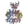
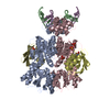
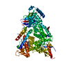
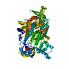
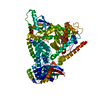
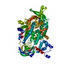
| Entry | Database: PDB / ID: 3oqn | ||||||
|---|---|---|---|---|---|---|---|
| Title | Structure of ccpa-hpr-ser46-p-gntr-down cre | ||||||
 Components Components |
| ||||||
 Keywords Keywords | TRANSCRIPTION/TRANSFERASE/DNA / pbp fold for ccpa / transcription / hpr-ser46p- / nucleoid / TRANSCRIPTION-TRANSFERASE-DNA complex | ||||||
| Function / homology |  Function and homology information Function and homology informationregulation of carbohydrate utilization / phosphoenolpyruvate-dependent sugar phosphotransferase system / DNA-binding transcription repressor activity / DNA-binding transcription activator activity / protein-DNA complex / transcription cis-regulatory region binding / DNA-binding transcription factor activity / negative regulation of DNA-templated transcription / regulation of DNA-templated transcription / positive regulation of DNA-templated transcription / cytoplasm Similarity search - Function | ||||||
| Biological species |  | ||||||
| Method |  X-RAY DIFFRACTION / SYNCHROTRON / MOLECULAR REPLACEMENT / Resolution: 3.3 Å X-RAY DIFFRACTION / SYNCHROTRON / MOLECULAR REPLACEMENT / Resolution: 3.3 Å | ||||||
 Authors Authors | Schumacher, M.A. / Sprehe, M. / Bartholomae, M. / Hillen, W. / Brennan, R.G. | ||||||
 Citation Citation | Journal: Nucleic Acids Res. / Year: 2011 Title: Structures of carbon catabolite protein A-(HPr-Ser46-P) bound to diverse catabolite response element sites reveal the basis for high-affinity binding to degenerate DNA operators. Authors: Schumacher, M.A. / Sprehe, M. / Bartholomae, M. / Hillen, W. / Brennan, R.G. | ||||||
| History |
|
- Structure visualization
| Structure viewer | Molecule: MolmilJmol/JSmol |
|---|
- Downloads & links
-Download
| PDBx/mmCIF format | 3oqn.cif.gz | 188.4 KB | Display | PDBx/mmCIF format |
|---|---|---|---|---|
| PDB format | pdb3oqn.ent.gz | 146 KB | Display | PDB format |
| PDBx/mmJSON format | 3oqn.json.gz | Tree view | PDBx/mmJSON format | |
| Others |  Other downloads Other downloads |
-Validation report
| Arichive directory | https://data.pdbj.org/pub/pdb/validation_reports/oq/3oqnftp://data.pdbj.org/pub/pdb/validation_reports/oq/3oqn | HTTPS FTP |
|---|
-Related structure data
| Related structure data |  3oqmC  3oqoC  2fepS C: citing same article ( S: Starting model for refinement |
|---|---|
| Similar structure data |
-Links
 PDBj
PDBj


- Assembly
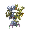
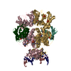
| Deposited unit | 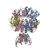
| ||||||||
|---|---|---|---|---|---|---|---|---|---|
| 1 |
| ||||||||
| Unit cell |
|
-Components
| #1: Protein | Mass: 37724.047 Da / Num. of mol.: 2 Source method: isolated from a genetically manipulated source Source: (gene. exp.) #2: Protein | Mass: 9147.117 Da / Num. of mol.: 2 Source method: isolated from a genetically manipulated source Source: (gene. exp.) References: UniProt: P08877, Transferases; Transferring phosphorus-containing groups; Protein-serine/threonine kinases #3: DNA chain | | Mass: 4922.216 Da / Num. of mol.: 1 / Source method: obtained synthetically / Source: (synth.) #4: DNA chain | | Mass: 4873.178 Da / Num. of mol.: 1 / Source method: obtained synthetically / Source: (synth.) Has protein modification | Y | |
|---|
-Experimental details
-Experiment
| Experiment | Method: X-RAY DIFFRACTION / Number of used crystals: 1 |
|---|
- Sample preparation
| Crystal | Density Matthews: 3.24 Å3/Da / Density % sol: 62.05 % |
|---|---|
| Crystal grow | Temperature: 298 K / Method: vapor diffusion, hanging drop / pH: 5 Details: peg 6000, citrate, pH 5.0, VAPOR DIFFUSION, HANGING DROP, temperature 298K |
-Data collection
| Diffraction | Mean temperature: 100 K |
|---|---|
| Diffraction source | Source: SYNCHROTRON / Site: ALS  / Beamline: 8.2.1 / Wavelength: 1 Å / Beamline: 8.2.1 / Wavelength: 1 Å |
| Detector | Type: ADSC QUANTUM 4 / Detector: CCD / Date: Dec 15, 2008 |
| Radiation | Monochromator: mirrors / Protocol: SINGLE WAVELENGTH / Monochromatic (M) / Laue (L): M / Scattering type: x-ray |
| Radiation wavelength | Wavelength: 1 Å / Relative weight: 1 |
| Reflection | Resolution: 3.3→87.7 Å / Num. all: 20642 / Num. obs: 19982 / % possible obs: 96.8 % / Observed criterion σ(F): 0 / Observed criterion σ(I): 0 |
| Reflection shell | Resolution: 3.3→3.56 Å / % possible all: 96.8 |
- Processing
| Software |
| ||||||||||||||||||||||||||||||||||||
|---|---|---|---|---|---|---|---|---|---|---|---|---|---|---|---|---|---|---|---|---|---|---|---|---|---|---|---|---|---|---|---|---|---|---|---|---|---|
| Refinement | Method to determine structure: MOLECULAR REPLACEMENT Starting model: 2FEP Resolution: 3.3→87.7 Å / Rfactor Rfree error: 0.008 / Data cutoff high absF: 2272253.13 / Data cutoff low absF: 0 / Isotropic thermal model: RESTRAINED / Cross valid method: THROUGHOUT / σ(F): 0 / Stereochemistry target values: Engh & Huber / Details: BULK SOLVENT MODEL USED
| ||||||||||||||||||||||||||||||||||||
| Solvent computation | Solvent model: FLAT MODEL / Bsol: 63.4853 Å2 / ksol: 0.35 e/Å3 | ||||||||||||||||||||||||||||||||||||
| Displacement parameters | Biso mean: 89 Å2
| ||||||||||||||||||||||||||||||||||||
| Refine analyze |
| ||||||||||||||||||||||||||||||||||||
| Refinement step | Cycle: LAST / Resolution: 3.3→87.7 Å
| ||||||||||||||||||||||||||||||||||||
| Refine LS restraints |
| ||||||||||||||||||||||||||||||||||||
| LS refinement shell | Resolution: 3.3→3.51 Å / Rfactor Rfree error: 0.025 / Total num. of bins used: 6
| ||||||||||||||||||||||||||||||||||||
| Xplor file |
|
