HDMs demethylate histones / Activated PKN1 stimulates transcription of AR (androgen receptor) regulated genes KLK2 and KLK3 / histone H3K36 demethylase activity / Recruitment and ATM-mediated phosphorylation of repair and signaling proteins at DNA double strand breaks / histone H3K9 demethylase activity / Estrogen-dependent gene expression / negative regulation of autophagy / positive regulation of transcription elongation by RNA polymerase II / DNA-binding transcription repressor activity, RNA polymerase II-specific / regulation of gene expression ...HDMs demethylate histones / Activated PKN1 stimulates transcription of AR (androgen receptor) regulated genes KLK2 and KLK3 / histone H3K36 demethylase activity / Recruitment and ATM-mediated phosphorylation of repair and signaling proteins at DNA double strand breaks / histone H3K9 demethylase activity / Estrogen-dependent gene expression / negative regulation of autophagy / positive regulation of transcription elongation by RNA polymerase II / DNA-binding transcription repressor activity, RNA polymerase II-specific / regulation of gene expression / sequence-specific DNA binding / chromatin remodeling / chromatin / negative regulation of transcription by RNA polymerase II / zinc ion binding / nucleus Similarity search - Function
JmjN domain / jmjN domain / JmjN domain profile. / Small domain found in the jumonji family of transcription factors / Cupin / JmjC domain, hydroxylase / Zinc finger, C2H2 type / A domain family that is part of the cupin metalloenzyme superfamily. / JmjC domain / JmjC domain profile. ...JmjN domain / jmjN domain / JmjN domain profile. / Small domain found in the jumonji family of transcription factors / Cupin / JmjC domain, hydroxylase / Zinc finger, C2H2 type / A domain family that is part of the cupin metalloenzyme superfamily. / JmjC domain / JmjC domain profile. / zinc finger / Zinc finger C2H2 type domain profile. / Zinc finger C2H2 superfamily / Zinc finger C2H2 type domain signature. / Zinc finger C2H2-type / Jelly Rolls / Sandwich / Mainly Beta Similarity search - Domain/homology
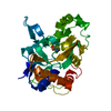
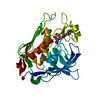
2-OXOGLUTARIC ACID / NICKEL (II) ION / DNA damage-responsive transcriptional repressor RPH1 Similarity search - Component
Biological species
Saccharomyces cerevisiae (brewer's yeast)
Method
X-RAY DIFFRACTION / SYNCHROTRON / SAD / Resolution: 2.2 Å
In the structure databanks used in Yorodumi, some data are registered as the other names, "COVID-19 virus" and "2019-nCoV". Here are the details of the virus and the list of structure data.
Jan 31, 2019. EMDB accession codes are about to change! (news from PDBe EMDB page)
EMDB accession codes are about to change! (news from PDBe EMDB page)
The allocation of 4 digits for EMDB accession codes will soon come to an end. Whilst these codes will remain in use, new EMDB accession codes will include an additional digit and will expand incrementally as the available range of codes is exhausted. The current 4-digit format prefixed with “EMD-” (i.e. EMD-XXXX) will advance to a 5-digit format (i.e. EMD-XXXXX), and so on. It is currently estimated that the 4-digit codes will be depleted around Spring 2019, at which point the 5-digit format will come into force.
The EM Navigator/Yorodumi systems omit the EMD- prefix.
Related info.:Q: What is EMD? / ID/Accession-code notation in Yorodumi/EM Navigator
Yorodumi is a browser for structure data from EMDB, PDB, SASBDB, etc.
This page is also the successor to EM Navigator detail page, and also detail information page/front-end page for Omokage search.
The word "yorodu" (or yorozu) is an old Japanese word meaning "ten thousand". "mi" (miru) is to see.
Related info.:EMDB / PDB / SASBDB / Comparison of 3 databanks / Yorodumi Search / Aug 31, 2016. New EM Navigator & Yorodumi / Yorodumi Papers / Jmol/JSmol / Function and homology information / Changes in new EM Navigator and Yorodumi
 Movie
Movie Controller

 Yorodumi
 Open data
 Basic information
 Components
 Keywords
 Function and homology information
 X-RAY DIFFRACTION /
  Authors
 Citation
 Structure visualization
 Downloads & links
 Other downloads
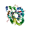


 PDBj
PDBj


 Assembly
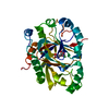


 Mass: 146.098 Da / Num. of mol.: 1 / Source method: obtained synthetically / Formula: C5H6O5
 Mass: 58.693 Da / Num. of mol.: 1 / Source method: obtained synthetically / Formula: Ni
 Mass: 18.015 Da / Num. of mol.: 226 / Source method: isolated from a natural source / Formula: H2O
 Sample preparation
 / Beamline: BL17U / Wavelength: 1 Å
 Processing