 Movie
Movie Controller


+ Open data

- Basic information
| Entry | Database: PDB / ID: 3oph | ||||||
|---|---|---|---|---|---|---|---|
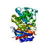
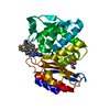
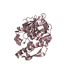
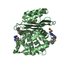
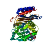
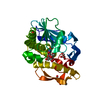
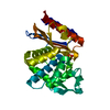
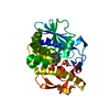
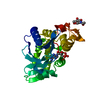
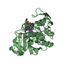
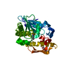
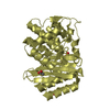
| Title | ESBL R164S mutant of SHV-1 beta-lactamase | ||||||
 Components Components | Beta-lactamase SHV-1 | ||||||
 Keywords Keywords | HYDROLASE / Class A beta-lactamase | ||||||
| Function / homology |  Function and homology information Function and homology informationbeta-lactam antibiotic catabolic process / beta-lactamase activity / beta-lactamase / response to antibiotic Similarity search - Function | ||||||
| Biological species |  Klebsiella pneumoniae (bacteria) Klebsiella pneumoniae (bacteria) | ||||||
| Method |  X-RAY DIFFRACTION / SYNCHROTRON / FOURIER SYNTHESIS / Resolution: 1.34 Å X-RAY DIFFRACTION / SYNCHROTRON / FOURIER SYNTHESIS / Resolution: 1.34 Å | ||||||
 Authors Authors | Sampson, J.M. / van den Akker, F. | ||||||
 Citation Citation | Journal: Antimicrob.Agents Chemother. / Year: 2011 Title: Ligand-dependent disorder of the Omega loop observed in extended-spectrum SHV-type beta-lactamase. Authors: Sampson, J.M. / Ke, W. / Bethel, C.R. / Pagadala, S.R. / Nottingham, M.D. / Bonomo, R.A. / Buynak, J.D. / van den Akker, F. | ||||||
| History |
|
- Structure visualization
| Structure viewer | Molecule: MolmilJmol/JSmol |
|---|
- Downloads & links
-Download
| PDBx/mmCIF format | 3oph.cif.gz | 77.2 KB | Display | PDBx/mmCIF format |
|---|---|---|---|---|
| PDB format | pdb3oph.ent.gz | 56.4 KB | Display | PDB format |
| PDBx/mmJSON format | 3oph.json.gz | Tree view | PDBx/mmJSON format | |
| Others |  Other downloads Other downloads |
-Validation report
| Arichive directory | https://data.pdbj.org/pub/pdb/validation_reports/op/3ophftp://data.pdbj.org/pub/pdb/validation_reports/op/3oph | HTTPS FTP |
|---|
-Related structure data
| Related structure data |  3oplC  3oppC  3oprC 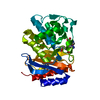 1vm1S C: citing same article ( S: Starting model for refinement |
|---|---|
| Similar structure data |


-Links
 PDBj
PDBj


- Assembly
| Deposited unit | 
| ||||||||
|---|---|---|---|---|---|---|---|---|---|
| 1 |
| ||||||||
| Unit cell |
| ||||||||
| Details | monomer in asymmetric unit |
-Components
| #1: Protein | Mass: 31189.875 Da / Num. of mol.: 1 / Mutation: R164S Source method: isolated from a genetically manipulated source Source: (gene. exp.) Klebsiella pneumoniae (bacteria) / Gene: bla, shv1 / Production host: | ||||||
|---|---|---|---|---|---|---|---|
| #2: Chemical |   Mass: 508.600 Da / Num. of mol.: 2 / Source method: obtained synthetically / Formula: C24H44O11 Mass: 508.600 Da / Num. of mol.: 2 / Source method: obtained synthetically / Formula: C24H44O11#3: Chemical | ChemComp-EPE / |   Mass: 238.305 Da / Num. of mol.: 1 / Source method: obtained synthetically / Formula: C8H18N2O4S / Comment: pH buffer*YM Mass: 238.305 Da / Num. of mol.: 1 / Source method: obtained synthetically / Formula: C8H18N2O4S / Comment: pH buffer*YM#4: Water | ChemComp-HOH / |  Mass: 18.015 Da / Num. of mol.: 341 / Source method: isolated from a natural source / Formula: H2O Mass: 18.015 Da / Num. of mol.: 341 / Source method: isolated from a natural source / Formula: H2OHas protein modification | Y | |
-Experimental details
-Experiment
| Experiment | Method: X-RAY DIFFRACTION |
|---|
- Sample preparation
| Crystal | Density Matthews: 1.82 Å3/Da / Density % sol: 32.56 % |
|---|---|
| Crystal grow | Temperature: 295 K / Method: vapor diffusion, sitting drop Details: 21-30% PEG 6000, 0.5mM cymal-6, 100mM HEPES buffer, pH 6.6-7.6, VAPOR DIFFUSION, SITTING DROP, temperature 295K PH range: 6.6-7.6 |
-Data collection
| Diffraction | Mean temperature: 100 K |
|---|---|
| Diffraction source | Source: SYNCHROTRON / Site: NSLS  / Beamline: X29A / Wavelength: 1.1 Å / Beamline: X29A / Wavelength: 1.1 Å |
| Detector | Type: ADSC QUANTUM 315r / Detector: CCD / Date: Oct 30, 2006 |
| Radiation | Monochromator: a vertically focusing mirror and a horizontally focusing monochromator Protocol: SINGLE WAVELENGTH / Monochromatic (M) / Laue (L): M / Scattering type: x-ray |
| Radiation wavelength | Wavelength: 1.1 Å / Relative weight: 1 |
| Reflection | Resolution: 1.34→50 Å / Num. all: 52100 / Num. obs: 51631 / % possible obs: 99.1 % / Observed criterion σ(F): 0 / Observed criterion σ(I): 0 / Redundancy: 3.5 % / Rmerge(I) obs: 0.061 / Net I/σ(I): 20.8 |
| Reflection shell | Resolution: 1.34→1.39 Å / Rmerge(I) obs: 0.234 / Mean I/σ(I) obs: 4.9 / % possible all: 94.6 |
- Processing
| Software |
| |||||||||||||||||||||||||||||||||||||||||||||||||||||||||||||||||
|---|---|---|---|---|---|---|---|---|---|---|---|---|---|---|---|---|---|---|---|---|---|---|---|---|---|---|---|---|---|---|---|---|---|---|---|---|---|---|---|---|---|---|---|---|---|---|---|---|---|---|---|---|---|---|---|---|---|---|---|---|---|---|---|---|---|---|
| Refinement | Method to determine structure: FOURIER SYNTHESIS Starting model: PDB entry 1VM1 Resolution: 1.34→31.79 Å / Cor.coef. Fo:Fc: 0.97 / Cor.coef. Fo:Fc free: 0.96 / SU B: 0.788 / SU ML: 0.034 / Cross valid method: THROUGHOUT / ESU R Free: 0.059 / Stereochemistry target values: MAXIMUM LIKELIHOOD / Details: HYDROGENS HAVE BEEN ADDED IN THE RIDING POSITIONS
| |||||||||||||||||||||||||||||||||||||||||||||||||||||||||||||||||
| Solvent computation | Ion probe radii: 0.8 Å / Shrinkage radii: 0.8 Å / VDW probe radii: 1.4 Å / Solvent model: MASK | |||||||||||||||||||||||||||||||||||||||||||||||||||||||||||||||||
| Displacement parameters | Biso mean: 15.205 Å2
| |||||||||||||||||||||||||||||||||||||||||||||||||||||||||||||||||
| Refinement step | Cycle: LAST / Resolution: 1.34→31.79 Å
| |||||||||||||||||||||||||||||||||||||||||||||||||||||||||||||||||
| Refine LS restraints |
| |||||||||||||||||||||||||||||||||||||||||||||||||||||||||||||||||
| LS refinement shell | Resolution: 1.34→1.376 Å / Total num. of bins used: 20
|
