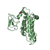
Entry Database : PDB / ID : 3onaTitle The SECRET domain in complex with CX3CL1 CX3CL1 protein Tumour necrosis factor receptor Keywords / / / / Function / homology Function Domain/homology Component
/ / / / / / / / / / / / / / / / / / / / / / / / / / / / / / / / / / / / / / / / / / / / / / / / / / / / / / / / / / / / / / / / / / / / / / / / / / / / / / / / / / / / / / / / / / / / / / / / / / / / / / / / / / / / / / / / / / / / / / / / / / / / / / Biological species Homo sapiens (human)Method / / / Resolution : 2.6 Å Authors Wang, X.Q. / Xue, X.G. / Wang, D.L. Journal : Plos Pathog. / Year : 2011Title : Structural basis of chemokine sequestration by CrmD, a poxvirus-encoded tumor necrosis factor receptorAuthors : Xue, X.G. / Lu, Q.Y. / Wei, H. / Wang, D.L. / Chen, D.W. / He, G.J. / Huang, L. / Wang, H.Z. / Wang, X.Q. History Deposition Aug 28, 2010 Deposition site / Processing site Revision 1.0 Aug 17, 2011 Provider / Type Revision 1.1 Jun 26, 2013 Group Revision 1.2 Nov 1, 2023 Group / Database references / Refinement descriptionCategory chem_comp_atom / chem_comp_bond ... chem_comp_atom / chem_comp_bond / database_2 / pdbx_initial_refinement_model / struct_ref_seq_dif Item / _database_2.pdbx_database_accession / _struct_ref_seq_dif.detailsRevision 1.3 Oct 30, 2024 Group / Category / pdbx_modification_feature
Show all Show less
 Movie
Movie Controller


 Open data
 Basic information
 Components
 Keywords
 Function and homology information
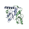
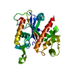
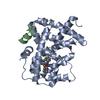
 Ectromelia virus
 Homo sapiens (human)
 X-RAY DIFFRACTION /
  Authors
 Citation
 Structure visualization
 Downloads & links
 Other downloads
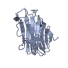


 PDBj
PDBj

 Assembly
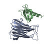

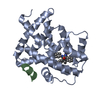
 Mass: 18.015 Da / Num. of mol.: 28 / Source method: isolated from a natural source / Formula: H2O
 Sample preparation
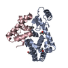
 / Beamline: BL17U / Wavelength: 0.97 Å
 Processing