 Movie
Movie Controller

[English] 日本語
 Yorodumi
- PDB-3ol0: Crystal structure of Monofoil-4P homo-trimer: de novo designed mo... -
+ Open data

- Basic information
| Entry | Database: PDB / ID: 3ol0 | ||||||
|---|---|---|---|---|---|---|---|
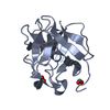
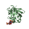
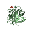
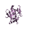
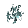
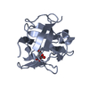
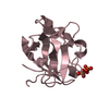
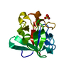
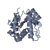
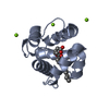
| Title | Crystal structure of Monofoil-4P homo-trimer: de novo designed monomer trefoil-fold sub-domain which forms homo-trimer assembly | ||||||
 Components Components | de novo designed monomer trefoil-fold sub-domain which forms homo-trimer assembly | ||||||
 Keywords Keywords | DE NOVO PROTEIN / beta-trefoil / trefoil-fold / synthetic protein / Function-competent only | ||||||
| Function / homology | SH3 type barrels. - #30 / SH3 type barrels. / Other non-globular / Special Function and homology information Function and homology information | ||||||
| Biological species | synthetic construct (others) | ||||||
| Method |  X-RAY DIFFRACTION / SYNCHROTRON / MOLECULAR REPLACEMENT / Resolution: 1.483 Å X-RAY DIFFRACTION / SYNCHROTRON / MOLECULAR REPLACEMENT / Resolution: 1.483 Å | ||||||
 Authors Authors | Lee, J. / Blaber, M. | ||||||
 Citation Citation | Journal: Proc.Natl.Acad.Sci.USA / Year: 2011 Title: Experimental support for the evolution of symmetric protein architecture from a simple peptide motif. Authors: Lee, J. / Blaber, M. | ||||||
| History |
|
- Structure visualization
| Structure viewer | Molecule: MolmilJmol/JSmol |
|---|
- Downloads & links
-Download
| PDBx/mmCIF format | 3ol0.cif.gz | 42.7 KB | Display | PDBx/mmCIF format |
|---|---|---|---|---|
| PDB format | pdb3ol0.ent.gz | 30.8 KB | Display | PDB format |
| PDBx/mmJSON format | 3ol0.json.gz | Tree view | PDBx/mmJSON format | |
| Others |  Other downloads Other downloads |
-Validation report
| Arichive directory | https://data.pdbj.org/pub/pdb/validation_reports/ol/3ol0ftp://data.pdbj.org/pub/pdb/validation_reports/ol/3ol0 | HTTPS FTP |
|---|
-Related structure data
| Related structure data |  3o49C  3o4aC  3o4bC 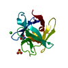 3o4cC 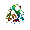 3o4dC  3ogfC C: citing same article ( |
|---|---|
| Similar structure data |


-Links
 PDBj
PDBj
- Assembly
| Deposited unit | 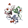
| ||||||||
|---|---|---|---|---|---|---|---|---|---|
| 1 |
| ||||||||
| Unit cell |
|
-Components
| #1: Protein/peptide | Mass: 5443.896 Da / Num. of mol.: 3 Source method: isolated from a genetically manipulated source Details: Synthetic sequence derived from human acidic fibroblast growth factor with a symmetric deconstruction method. The protein produced by this sequence forms a monomer trefoil-fold sub-domain ...Details: Synthetic sequence derived from human acidic fibroblast growth factor with a symmetric deconstruction method. The protein produced by this sequence forms a monomer trefoil-fold sub-domain and exists as a homo-trimer assembly adopting a beta-trefoil architecture. Source: (gene. exp.) synthetic construct (others) / Production host:  #2: Chemical | ChemComp-SO4 /   Mass: 96.063 Da / Num. of mol.: 5 / Source method: obtained synthetically / Formula: SO4 Mass: 96.063 Da / Num. of mol.: 5 / Source method: obtained synthetically / Formula: SO4#3: Water | ChemComp-HOH / |  Mass: 18.015 Da / Num. of mol.: 203 / Source method: isolated from a natural source / Formula: H2O Mass: 18.015 Da / Num. of mol.: 203 / Source method: isolated from a natural source / Formula: H2O |
|---|
-Experimental details
-Experiment
| Experiment | Method: X-RAY DIFFRACTION / Number of used crystals: 2 |
|---|
- Sample preparation
| Crystal | Density Matthews: 2.67 Å3/Da / Density % sol: 53.94 % |
|---|---|
| Crystal grow | Temperature: 298 K / Method: vapor diffusion, hanging drop / pH: 5.5 Details: 2M AMMONIUM SULFATE, 0.1M NA CITRATE, 15MG/ML PROTEIN CONCENTRATION, Ph 5.5, VAPOR DIFFUSION, HANGING DROP, TEMPERATURE 298K |
-Data collection
| Diffraction | Mean temperature: 100 K |
|---|---|
| Diffraction source | Source: SYNCHROTRON / Site: NSLS  / Beamline: X29A / Wavelength: 1.075 Å / Beamline: X29A / Wavelength: 1.075 Å |
| Detector | Type: ADSC QUANTUM 315 / Detector: CCD / Date: Aug 23, 2010 |
| Radiation | Monochromator: SI / Protocol: SINGLE WAVELENGTH / Monochromatic (M) / Laue (L): M / Scattering type: x-ray |
| Radiation wavelength | Wavelength: 1.075 Å / Relative weight: 1 |
| Reflection | Resolution: 1.48→50 Å / Num. all: 29633 / Num. obs: 29337 / % possible obs: 99 % / Observed criterion σ(F): 3 / Observed criterion σ(I): 3 / Redundancy: 19.4 % / Rmerge(I) obs: 0.069 / Net I/σ(I): 82.4 |
| Reflection shell | Resolution: 1.48→1.51 Å / Redundancy: 16.6 % / Rmerge(I) obs: 0.358 / Mean I/σ(I) obs: 8.38 / Num. unique all: 1434 / % possible all: 98.8 |
- Processing
| Software |
| |||||||||||||||||||||||||||||||||||||||||||||||||||||||||||||||||||||||||||||
|---|---|---|---|---|---|---|---|---|---|---|---|---|---|---|---|---|---|---|---|---|---|---|---|---|---|---|---|---|---|---|---|---|---|---|---|---|---|---|---|---|---|---|---|---|---|---|---|---|---|---|---|---|---|---|---|---|---|---|---|---|---|---|---|---|---|---|---|---|---|---|---|---|---|---|---|---|---|---|
| Refinement | Method to determine structure: MOLECULAR REPLACEMENT / Resolution: 1.483→41.517 Å / SU ML: 0.14 / σ(F): 4.48 / Stereochemistry target values: ML
| |||||||||||||||||||||||||||||||||||||||||||||||||||||||||||||||||||||||||||||
| Solvent computation | Shrinkage radii: 0.9 Å / VDW probe radii: 1.11 Å / Solvent model: FLAT BULK SOLVENT MODEL / Bsol: 37.175 Å2 / ksol: 0.376 e/Å3 | |||||||||||||||||||||||||||||||||||||||||||||||||||||||||||||||||||||||||||||
| Displacement parameters |
| |||||||||||||||||||||||||||||||||||||||||||||||||||||||||||||||||||||||||||||
| Refinement step | Cycle: LAST / Resolution: 1.483→41.517 Å
| |||||||||||||||||||||||||||||||||||||||||||||||||||||||||||||||||||||||||||||
| Refine LS restraints |
| |||||||||||||||||||||||||||||||||||||||||||||||||||||||||||||||||||||||||||||
| LS refinement shell | Refine-ID: X-RAY DIFFRACTION
|
