 Movie
Movie Controller

[English] 日本語
 Yorodumi
- PDB-3oiz: Crystal structure of antisigma-factor antagonist, STAS domain fro... -
+ Open data

- Basic information
| Entry | Database: PDB / ID: 3oiz | ||||||
|---|---|---|---|---|---|---|---|
| Title | Crystal structure of antisigma-factor antagonist, STAS domain from Rhodobacter sphaeroides | ||||||
 Components Components | Antisigma-factor antagonist, STAS | ||||||
 Keywords Keywords | MEMBRANE PROTEIN / PSI-2 / Midwest Center for Structural Genomics / Protein Structure Initiative / MCSG / STAS domain | ||||||
| Function / homology |  Function and homology information Function and homology information | ||||||
| Biological species |  Rhodobacter sphaeroides (bacteria) Rhodobacter sphaeroides (bacteria) | ||||||
| Method |  X-RAY DIFFRACTION / SYNCHROTRON / SAD / Resolution: 1.65 Å X-RAY DIFFRACTION / SYNCHROTRON / SAD / Resolution: 1.65 Å | ||||||
 Authors Authors | Chang, C. / Marshall, N. / Freeman, L. / Joachimiak, A. / Midwest Center for Structural Genomics (MCSG) | ||||||
 Citation Citation | Journal: To be Published Title: Crystal structure of antisigma-factor antagonist, STAS domain from Rhodobacter sphaeroides Authors: Chang, C. / Marshall, N. / Freeman, L. / Joachimiak, A. | ||||||
| History |
|
- Structure visualization
| Structure viewer | Molecule: MolmilJmol/JSmol |
|---|
- Downloads & links
-Download
| PDBx/mmCIF format | 3oiz.cif.gz | 56.3 KB | Display | PDBx/mmCIF format |
|---|---|---|---|---|
| PDB format | pdb3oiz.ent.gz | 41.9 KB | Display | PDB format |
| PDBx/mmJSON format | 3oiz.json.gz | Tree view | PDBx/mmJSON format | |
| Others |  Other downloads Other downloads |
-Validation report
| Arichive directory | https://data.pdbj.org/pub/pdb/validation_reports/oi/3oizftp://data.pdbj.org/pub/pdb/validation_reports/oi/3oiz | HTTPS FTP |
|---|
-Related structure data
| Similar structure data | |
|---|---|
| Other databases |


-Links
 PDBj
PDBj
- Assembly
| Deposited unit | 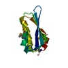
| ||||||||
|---|---|---|---|---|---|---|---|---|---|
| 1 |
| ||||||||
| Unit cell |
|
-Components
| #1: Protein | Mass: 11442.281 Da / Num. of mol.: 1 Source method: isolated from a genetically manipulated source Source: (gene. exp.) Rhodobacter sphaeroides (bacteria) / Strain: ATCC 17023 / 2.4.1 / NCIB 8253 / DSM 158 / Gene: RHOS4_40800, RSP_4197 / Plasmid: pMCSG19 / Production host: |
|---|---|
| #2: Water | ChemComp-HOH /  Mass: 18.015 Da / Num. of mol.: 110 / Source method: isolated from a natural source / Formula: H2O Mass: 18.015 Da / Num. of mol.: 110 / Source method: isolated from a natural source / Formula: H2O |
| Has protein modification | Y |
-Experimental details
-Experiment
| Experiment | Method: X-RAY DIFFRACTION / Number of used crystals: 1 |
|---|
- Sample preparation
| Crystal | Density Matthews: 2.07 Å3/Da / Density % sol: 40.55 % |
|---|---|
| Crystal grow | Temperature: 297 K / Method: vapor diffusion, sitting drop / pH: 6 Details: 0.2M Calcium acetate, 0.1M MES, 10% iso-propanol, pH 6.0, VAPOR DIFFUSION, SITTING DROP, temperature 297K |
-Data collection
| Diffraction | Mean temperature: 100 K |
|---|---|
| Diffraction source | Source: SYNCHROTRON / Site: APS  / Beamline: 19-ID / Wavelength: 0.97935 Å / Beamline: 19-ID / Wavelength: 0.97935 Å |
| Detector | Type: ADSC QUANTUM 315r / Detector: CCD / Date: Jun 7, 2010 |
| Radiation | Monochromator: Si(111) double crystal / Protocol: SINGLE WAVELENGTH / Monochromatic (M) / Laue (L): M / Scattering type: x-ray |
| Radiation wavelength | Wavelength: 0.97935 Å / Relative weight: 1 |
| Reflection | Resolution: 1.65→50 Å / Num. all: 12087 / Num. obs: 12047 / % possible obs: 99.7 % / Observed criterion σ(I): -3 / Redundancy: 10.5 % / Biso Wilson estimate: 24.9 Å2 / Rmerge(I) obs: 0.097 / Net I/σ(I): 62.2 |
| Reflection shell | Resolution: 1.65→1.68 Å / Redundancy: 4.9 % / Rmerge(I) obs: 0.507 / Mean I/σ(I) obs: 2.9 / Num. unique all: 582 / % possible all: 99.7 |
- Processing
| Software |
| ||||||||||||||||||||||||||||||||||||||||||||||||||||||||||||||||||||||
|---|---|---|---|---|---|---|---|---|---|---|---|---|---|---|---|---|---|---|---|---|---|---|---|---|---|---|---|---|---|---|---|---|---|---|---|---|---|---|---|---|---|---|---|---|---|---|---|---|---|---|---|---|---|---|---|---|---|---|---|---|---|---|---|---|---|---|---|---|---|---|---|
| Refinement | Method to determine structure: SAD / Resolution: 1.65→50 Å / Cor.coef. Fo:Fc: 0.969 / Cor.coef. Fo:Fc free: 0.954 / Occupancy max: 1 / Occupancy min: 0.5 / SU B: 4.192 / SU ML: 0.065 / Cross valid method: THROUGHOUT / σ(F): 0 / ESU R Free: 0.101 Stereochemistry target values: MAXIMUM LIKELIHOOD WITH PHASES Details: HYDROGENS HAVE BEEN ADDED IN THE RIDING POSITIONS U VALUES : WITH TLS ADDED
| ||||||||||||||||||||||||||||||||||||||||||||||||||||||||||||||||||||||
| Solvent computation | Ion probe radii: 0.8 Å / Shrinkage radii: 0.8 Å / VDW probe radii: 1.4 Å / Solvent model: MASK | ||||||||||||||||||||||||||||||||||||||||||||||||||||||||||||||||||||||
| Displacement parameters | Biso max: 80.33 Å2 / Biso mean: 30.2107 Å2 / Biso min: 18.35 Å2
| ||||||||||||||||||||||||||||||||||||||||||||||||||||||||||||||||||||||
| Refinement step | Cycle: LAST / Resolution: 1.65→50 Å
| ||||||||||||||||||||||||||||||||||||||||||||||||||||||||||||||||||||||
| Refine LS restraints |
| ||||||||||||||||||||||||||||||||||||||||||||||||||||||||||||||||||||||
| LS refinement shell | Resolution: 1.648→1.691 Å / Total num. of bins used: 20
| ||||||||||||||||||||||||||||||||||||||||||||||||||||||||||||||||||||||
| Refinement TLS params. | Method: refined / Origin x: 35.8237 Å / Origin y: 17.1956 Å / Origin z: 16.8824 Å
|
