Resolution: 3.011→28.89 Å / SU ML: 0.44 / σ(F): 1.97 / Phase error: 33.22 / Stereochemistry target values: ML Details: AUTHORS HAVE APPROVED THIS ENTRY WITH REPORTED R VALUE AS 0.23 WHILE CALCULATED R VALUE IS 0.31. THE DEPOSITED COORDINATES WERE FURTHER MODIFIED IN COOT AFTER FINAL ROUND OF PHENIX REFINEMENT.
Rfactor
Num. reflection
% reflection
Selection details
Rfree
0.3053
1368
5 %
random
Rwork
0.2295
-
-
-
obs
0.2334
27375
98.04 %
-
all
-
27410
-
-
Solvent computation
Shrinkage radii: 0.9 Å / VDW probe radii: 1.11 Å / Solvent model: FLAT BULK SOLVENT MODEL / Bsol: 48.835 Å2 / ksol: 0.29 e/Å3
Displacement parameters
Biso mean: 61.8 Å2
Baniso -1
Baniso -2
Baniso -3
1-
-8.2886 Å2
4.2899 Å2
-5.1891 Å2
2-
-
18.7583 Å2
-5.878 Å2
3-
-
-
-10.4697 Å2
Refinement step
Cycle: LAST / Resolution: 3.011→28.89 Å
Protein
Nucleic acid
Ligand
Solvent
Total
Num. atoms
7246
0
0
0
7246
Refine LS restraints
Refine-ID
Type
Dev ideal
Number
X-RAY DIFFRACTION
f_bond_d
0.009
7426
X-RAY DIFFRACTION
f_angle_d
1.387
10068
X-RAY DIFFRACTION
f_dihedral_angle_d
18.11
2603
X-RAY DIFFRACTION
f_chiral_restr
0.09
1061
X-RAY DIFFRACTION
f_plane_restr
0.005
1323
LS refinement shell
Resolution (Å)
Rfactor Rfree
Num. reflection Rfree
Rfactor Rwork
Num. reflection Rwork
Refine-ID
% reflection obs (%)
3.0113-3.1188
0.3784
128
0.2699
2526
X-RAY DIFFRACTION
94
3.1188-3.2435
0.3584
125
0.2427
2607
X-RAY DIFFRACTION
99
3.2435-3.3909
0.3001
123
0.2269
2601
X-RAY DIFFRACTION
98
3.3909-3.5693
0.2785
130
0.2083
2645
X-RAY DIFFRACTION
99
3.5693-3.7925
0.2803
156
0.2021
2612
X-RAY DIFFRACTION
99
3.7925-4.0846
0.2941
119
0.2079
2631
X-RAY DIFFRACTION
99
4.0846-4.4943
0.2464
146
0.1897
2609
X-RAY DIFFRACTION
99
4.4943-5.1414
0.2589
125
0.1878
2627
X-RAY DIFFRACTION
99
5.1414-6.4657
0.3307
158
0.2575
2599
X-RAY DIFFRACTION
98
6.4657-28.8916
0.33
158
0.2671
2550
X-RAY DIFFRACTION
97
+
About Yorodumi
-
News
-
Feb 9, 2022. New format data for meta-information of EMDB entries
New format data for meta-information of EMDB entries
Version 3 of the EMDB header file is now the official format.
The previous official version 1.9 will be removed from the archive.
In the structure databanks used in Yorodumi, some data are registered as the other names, "COVID-19 virus" and "2019-nCoV". Here are the details of the virus and the list of structure data.
Jan 31, 2019. EMDB accession codes are about to change! (news from PDBe EMDB page)
EMDB accession codes are about to change! (news from PDBe EMDB page)
The allocation of 4 digits for EMDB accession codes will soon come to an end. Whilst these codes will remain in use, new EMDB accession codes will include an additional digit and will expand incrementally as the available range of codes is exhausted. The current 4-digit format prefixed with “EMD-” (i.e. EMD-XXXX) will advance to a 5-digit format (i.e. EMD-XXXXX), and so on. It is currently estimated that the 4-digit codes will be depleted around Spring 2019, at which point the 5-digit format will come into force.
The EM Navigator/Yorodumi systems omit the EMD- prefix.
Related info.:Q: What is EMD? / ID/Accession-code notation in Yorodumi/EM Navigator
Yorodumi is a browser for structure data from EMDB, PDB, SASBDB, etc.
This page is also the successor to EM Navigator detail page, and also detail information page/front-end page for Omokage search.
The word "yorodu" (or yorozu) is an old Japanese word meaning "ten thousand". "mi" (miru) is to see.
Related info.:EMDB / PDB / SASBDB / Comparison of 3 databanks / Yorodumi Search / Aug 31, 2016. New EM Navigator & Yorodumi / Yorodumi Papers / Jmol/JSmol / Function and homology information / Changes in new EM Navigator and Yorodumi
 Movie
Movie Controller


 Open data
 Basic information
 Components
 Keywords
 Function and homology information
 X-RAY DIFFRACTION /
  Authors
 Citation
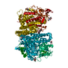
 Structure visualization
 Downloads & links
 Other downloads


 PDBj
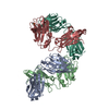
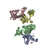
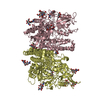
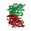
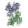
PDBj Assembly


 Sample preparation
 / Beamline: X25 / Wavelength: 1 Å
 Processing