 Movie
Movie Controller


+ Open data

- Basic information
| Entry | Database: PDB / ID: 3ogd | ||||||
|---|---|---|---|---|---|---|---|
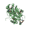
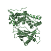
| Title | AlkA Undamaged DNA Complex: Interrogation of a G*:C base pair | ||||||
 Components Components |
| ||||||
 Keywords Keywords | HYDROLASE/DNA / helix-hairpin-helix / DNA repair / Alkylation / HYDROLASE-DNA complex | ||||||
| Function / homology |  Function and homology information Function and homology informationDNA-7-methylguanine glycosylase activity / alkylated DNA binding / DNA-3-methyladenine glycosylase activity / alkylbase DNA N-glycosylase activity / DNA-3-methyladenine glycosylase II / base-excision repair, AP site formation / DNA alkylation repair / base-excision repair / DNA repair / DNA damage response / cytoplasm Similarity search - Function | ||||||
| Biological species |  | ||||||
| Method |  X-RAY DIFFRACTION / SYNCHROTRON / MOLECULAR REPLACEMENT / Resolution: 2.8 Å X-RAY DIFFRACTION / SYNCHROTRON / MOLECULAR REPLACEMENT / Resolution: 2.8 Å | ||||||
 Authors Authors | Bowman, B.R. / Lee, S. / Wang, S. / Verdine, G.L. | ||||||
 Citation Citation | Journal: J.Biol.Chem. / Year: 2010 Title: Structure of Escherichia coli AlkA in Complex with Undamaged DNA. Authors: Bowman, B.R. / Lee, S. / Wang, S. / Verdine, G.L. | ||||||
| History |
|
- Structure visualization
| Structure viewer | Molecule: MolmilJmol/JSmol |
|---|
- Downloads & links
-Download
| PDBx/mmCIF format | 3ogd.cif.gz | 82.8 KB | Display | PDBx/mmCIF format |
|---|---|---|---|---|
| PDB format | pdb3ogd.ent.gz | 58.2 KB | Display | PDB format |
| PDBx/mmJSON format | 3ogd.json.gz | Tree view | PDBx/mmJSON format | |
| Others |  Other downloads Other downloads |
-Validation report
| Arichive directory | https://data.pdbj.org/pub/pdb/validation_reports/og/3ogdftp://data.pdbj.org/pub/pdb/validation_reports/og/3ogd | HTTPS FTP |
|---|
-Related structure data
| Related structure data | 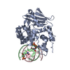 3oh6C 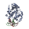 3oh9C  1dizS C: citing same article ( S: Starting model for refinement |
|---|---|
| Similar structure data |


-Links
 PDBj
PDBj- Assembly
| Deposited unit | 
| ||||||||
|---|---|---|---|---|---|---|---|---|---|
| 1 |
| ||||||||
| Unit cell |
|
-Components
| #1: Protein | Mass: 32134.967 Da / Num. of mol.: 1 / Mutation: L125C Source method: isolated from a genetically manipulated source Source: (gene. exp.) References: UniProt: P04395, DNA-3-methyladenine glycosylase II |
|---|---|
| #2: DNA chain | Mass: 2755.823 Da / Num. of mol.: 1 / Source method: obtained synthetically |
| #3: DNA chain | Mass: 2845.539 Da / Num. of mol.: 1 / Source method: obtained synthetically |
-Experimental details
-Experiment
| Experiment | Method: X-RAY DIFFRACTION / Number of used crystals: 1 |
|---|
- Sample preparation
| Crystal | Density Matthews: 3.08 Å3/Da / Density % sol: 60.07 % |
|---|---|
| Crystal grow | Temperature: 298 K / Method: vapor diffusion, sitting drop Details: 25-29% peg3350, 100mM Bis-Tris 6.0-6.6, 200mM LiSo, 3% 6-aminocaproic acid, pH 6.0-6.5, VAPOR DIFFUSION, SITTING DROP, temperature 298K PH range: 6.0-6.5 |
-Data collection
| Diffraction | Mean temperature: 77 K | ||||||||||||||||||||||||||||||||||||||||||||||||||||||||||||||||||
|---|---|---|---|---|---|---|---|---|---|---|---|---|---|---|---|---|---|---|---|---|---|---|---|---|---|---|---|---|---|---|---|---|---|---|---|---|---|---|---|---|---|---|---|---|---|---|---|---|---|---|---|---|---|---|---|---|---|---|---|---|---|---|---|---|---|---|---|
| Diffraction source | Source: SYNCHROTRON / Site: APS  / Beamline: 24-ID-C / Wavelength: 0.9197 Å / Beamline: 24-ID-C / Wavelength: 0.9197 Å | ||||||||||||||||||||||||||||||||||||||||||||||||||||||||||||||||||
| Detector | Type: ADSC QUANTUM 4 / Detector: CCD / Date: Nov 25, 2008 | ||||||||||||||||||||||||||||||||||||||||||||||||||||||||||||||||||
| Radiation | Scattering type: x-ray | ||||||||||||||||||||||||||||||||||||||||||||||||||||||||||||||||||
| Radiation wavelength | Wavelength: 0.9197 Å / Relative weight: 1 | ||||||||||||||||||||||||||||||||||||||||||||||||||||||||||||||||||
| Reflection | Resolution: 2.779→50 Å / Num. obs: 12042 / % possible obs: 99.5 % / Redundancy: 7.6 % / Rmerge(I) obs: 0.072 / Χ2: 1.349 / Net I/σ(I): 12.4 | ||||||||||||||||||||||||||||||||||||||||||||||||||||||||||||||||||
| Reflection shell |
|
- Processing
| Software |
| ||||||||||||||||||||||||||||||||||||||||||||||||||||||||
|---|---|---|---|---|---|---|---|---|---|---|---|---|---|---|---|---|---|---|---|---|---|---|---|---|---|---|---|---|---|---|---|---|---|---|---|---|---|---|---|---|---|---|---|---|---|---|---|---|---|---|---|---|---|---|---|---|---|
| Refinement | Method to determine structure: MOLECULAR REPLACEMENT Starting model: 1DIZ Resolution: 2.8→44.177 Å / Occupancy max: 1 / Occupancy min: 1 / FOM work R set: 0.7759 / SU ML: 2.58 / σ(F): 0.16 / Stereochemistry target values: ML
| ||||||||||||||||||||||||||||||||||||||||||||||||||||||||
| Solvent computation | Shrinkage radii: 0.9 Å / VDW probe radii: 1.11 Å / Solvent model: FLAT BULK SOLVENT MODEL / Bsol: 50.543 Å2 / ksol: 0.286 e/Å3 | ||||||||||||||||||||||||||||||||||||||||||||||||||||||||
| Displacement parameters | Biso max: 207 Å2 / Biso mean: 102.6048 Å2 / Biso min: 60.54 Å2
| ||||||||||||||||||||||||||||||||||||||||||||||||||||||||
| Refinement step | Cycle: LAST / Resolution: 2.8→44.177 Å
| ||||||||||||||||||||||||||||||||||||||||||||||||||||||||
| Refine LS restraints |
| ||||||||||||||||||||||||||||||||||||||||||||||||||||||||
| LS refinement shell | Refine-ID: X-RAY DIFFRACTION / Total num. of bins used: 7
|
