 Movie
Movie Controller

[English] 日本語
 Yorodumi
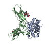
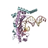
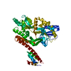
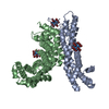
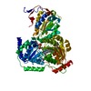
- PDB-3og6: The crystal structure of human interferon lambda 1 complexed with... -
+ Open data

- Basic information
| Entry | Database: PDB / ID: 3og6 | |||||||||
|---|---|---|---|---|---|---|---|---|---|---|
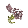
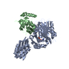
| Title | The crystal structure of human interferon lambda 1 complexed with its high affinity receptor in space group P212121 | |||||||||
 Components Components |
| |||||||||
 Keywords Keywords | CYTOKINE/CYTOKINE RECEPTOR / Helical bundle / fibronectin type III domain / beta-sandwich / Cytokine signaling / Membrane / CYTOKINE-CYTOKINE RECEPTOR complex | |||||||||
| Function / homology |  Function and homology information Function and homology informationinterleukin-28 receptor binding / negative regulation of memory T cell differentiation / positive regulation of MHC class I biosynthetic process / response to type III interferon / interleukin-28 receptor complex / negative regulation of type 2 immune response / negative regulation of interleukin-5 production / negative regulation of interleukin-13 production / mucosal immune response / negative regulation of T cell differentiation ...interleukin-28 receptor binding / negative regulation of memory T cell differentiation / positive regulation of MHC class I biosynthetic process / response to type III interferon / interleukin-28 receptor complex / negative regulation of type 2 immune response / negative regulation of interleukin-5 production / negative regulation of interleukin-13 production / mucosal immune response / negative regulation of T cell differentiation / positive regulation of cellular respiration / type III interferon-mediated signaling pathway / regulation of defense response to virus by host / cytokine receptor activity / Other interleukin signaling / positive regulation of tyrosine phosphorylation of STAT protein / Interleukin-20 family signaling / cell surface receptor signaling pathway via JAK-STAT / cytokine activity / positive regulation of receptor signaling pathway via JAK-STAT / cellular response to virus / positive regulation of immune response / positive regulation of type II interferon production / cytokine-mediated signaling pathway / defense response to virus / signaling receptor binding / negative regulation of cell population proliferation / innate immune response / negative regulation of DNA-templated transcription / positive regulation of DNA-templated transcription / : / extracellular region / membrane / plasma membrane Similarity search - Function | |||||||||
| Biological species |  Homo sapiens (human) Homo sapiens (human) | |||||||||
| Method |  X-RAY DIFFRACTION / SYNCHROTRON / MOLECULAR REPLACEMENT / molecular replacement / Resolution: 2.097 Å X-RAY DIFFRACTION / SYNCHROTRON / MOLECULAR REPLACEMENT / molecular replacement / Resolution: 2.097 Å | |||||||||
 Authors Authors | Miknis, Z.J. / Magracheva, E. / Lei, W. / Zdanov, A. / Kotenko, S.V. / Wlodawer, A. | |||||||||
 Citation Citation | Journal: J.Mol.Biol. / Year: 2010 Title: Crystal structure of the complex of human interferon-lambda1 with its high affinity receptor interferon-lambdaR1. Authors: Miknis, Z.J. / Magracheva, E. / Li, W. / Zdanov, A. / Kotenko, S.V. / Wlodawer, A. | |||||||||
| History |
|
- Structure visualization
| Structure viewer | Molecule: MolmilJmol/JSmol |
|---|
- Downloads & links
-Download
| PDBx/mmCIF format | 3og6.cif.gz | 166.6 KB | Display | PDBx/mmCIF format |
|---|---|---|---|---|
| PDB format | pdb3og6.ent.gz | 130.1 KB | Display | PDB format |
| PDBx/mmJSON format | 3og6.json.gz | Tree view | PDBx/mmJSON format | |
| Others |  Other downloads Other downloads |
-Validation report
| Arichive directory | https://data.pdbj.org/pub/pdb/validation_reports/og/3og6ftp://data.pdbj.org/pub/pdb/validation_reports/og/3og6 | HTTPS FTP |
|---|
-Related structure data
| Related structure data |  3og4C 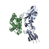 3g9vS 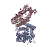 3hhcS S: Starting model for refinement C: citing same article ( |
|---|---|
| Similar structure data |

-Links
 PDBj
PDBj


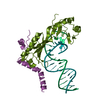
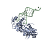
- Assembly
| Deposited unit | 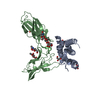
| ||||||||
|---|---|---|---|---|---|---|---|---|---|
| 1 |
| ||||||||
| Unit cell |
|
-Components
-Protein , 2 types, 2 molecules AB
| #1: Protein | Mass: 22000.043 Da / Num. of mol.: 1 / Mutation: G1A, T10P Source method: isolated from a genetically manipulated source Source: (gene. exp.) Homo sapiens (human) / Gene: IFNL1, IL29, ZCYTO21 / Plasmid: pMT/BiP/V5-His / Production host:  |
|---|---|
| #2: Protein | Mass: 25320.770 Da / Num. of mol.: 1 / Fragment: Extracellular domain (UNP residue 19-226) Source method: isolated from a genetically manipulated source Source: (gene. exp.) Homo sapiens (human) / Gene: hCG_1982865, IL28RA, RP11-10N16.1-001 / Plasmid: pMT/BiP/V5-His / Production host: |
-Sugars , 2 types, 3 molecules 
| #3: Polysaccharide | beta-D-mannopyranose-(1-4)-2-acetamido-2-deoxy-beta-D-glucopyranose-(1-4)-2-acetamido-2-deoxy-beta- ...beta-D-mannopyranose-(1-4)-2-acetamido-2-deoxy-beta-D-glucopyranose-(1-4)-2-acetamido-2-deoxy-beta-D-glucopyranose Source method: isolated from a genetically manipulated source |
|---|---|
| #5: Sugar |  Type: D-saccharide, beta linking / Mass: 221.208 Da / Num. of mol.: 2 Type: D-saccharide, beta linking / Mass: 221.208 Da / Num. of mol.: 2Source method: isolated from a genetically manipulated source Formula: C8H15NO6 |
-Non-polymers , 2 types, 270 molecules 

| #4: Chemical | ChemComp-GOL /  Mass: 92.094 Da / Num. of mol.: 4 / Source method: obtained synthetically / Formula: C3H8O3 Mass: 92.094 Da / Num. of mol.: 4 / Source method: obtained synthetically / Formula: C3H8O3#6: Water | ChemComp-HOH / | Mass: 18.015 Da / Num. of mol.: 266 / Source method: isolated from a natural source / Formula: H2O |
|---|
-Details
| Has protein modification | Y |
|---|
-Experimental details
-Experiment
| Experiment | Method: X-RAY DIFFRACTION / Number of used crystals: 1 |
|---|
- Sample preparation
| Crystal | Density Matthews: 3.43 Å3/Da / Density % sol: 64.14 % |
|---|---|
| Crystal grow | Temperature: 298 K / Method: vapor diffusion, hanging drop / pH: 7.9 Details: 20% MPEG 2000, 100 mM Tris-HCl pH 7.9, 200 mM trimethyamine N-oxide dehydrate, VAPOR DIFFUSION, HANGING DROP, temperature 298K |
-Data collection
| Diffraction | Mean temperature: 100 K | |||||||||||||||||||||||||||||||||||||||||||||||||||||||||||||||||||||||||||||
|---|---|---|---|---|---|---|---|---|---|---|---|---|---|---|---|---|---|---|---|---|---|---|---|---|---|---|---|---|---|---|---|---|---|---|---|---|---|---|---|---|---|---|---|---|---|---|---|---|---|---|---|---|---|---|---|---|---|---|---|---|---|---|---|---|---|---|---|---|---|---|---|---|---|---|---|---|---|---|
| Diffraction source | Source: SYNCHROTRON / Site: APS  / Beamline: 22-ID / Wavelength: 1 Å / Beamline: 22-ID / Wavelength: 1 Å | |||||||||||||||||||||||||||||||||||||||||||||||||||||||||||||||||||||||||||||
| Detector | Type: MARMOSAIC 300 mm CCD / Detector: CCD / Date: Jun 30, 2009 Details: Rosenbaum-Rock monochromator high-resolution double-crystal Si(220) sagittal focusing, Rosenbaum-Rock vertical focusing mirror | |||||||||||||||||||||||||||||||||||||||||||||||||||||||||||||||||||||||||||||
| Radiation | Protocol: SINGLE WAVELENGTH / Monochromatic (M) / Laue (L): M / Scattering type: x-ray | |||||||||||||||||||||||||||||||||||||||||||||||||||||||||||||||||||||||||||||
| Radiation wavelength | Wavelength: 1 Å / Relative weight: 1 | |||||||||||||||||||||||||||||||||||||||||||||||||||||||||||||||||||||||||||||
| Reflection | Redundancy: 6.8 % / Av σ(I) over netI: 23.64 / Number: 258890 / Rmerge(I) obs: 0.07 / Χ2: 0.92 / D res high: 2.1 Å / D res low: 30 Å / Num. obs: 37819 / % possible obs: 97.1 | |||||||||||||||||||||||||||||||||||||||||||||||||||||||||||||||||||||||||||||
| Diffraction reflection shell |
| |||||||||||||||||||||||||||||||||||||||||||||||||||||||||||||||||||||||||||||
| Reflection | Resolution: 2.1→30 Å / Num. all: 38948 / Num. obs: 37819 / % possible obs: 97.1 % / Redundancy: 6.8 % / Biso Wilson estimate: 37.63 Å2 / Rmerge(I) obs: 0.07 / Χ2: 0.919 / Net I/σ(I): 10.5 | |||||||||||||||||||||||||||||||||||||||||||||||||||||||||||||||||||||||||||||
| Reflection shell |
|
-Phasing
| Phasing | Method: molecular replacement |
|---|
- Processing
| Software |
| |||||||||||||||||||||||||||||||||||||||||||||||||||||||||||||||||||||||||||
|---|---|---|---|---|---|---|---|---|---|---|---|---|---|---|---|---|---|---|---|---|---|---|---|---|---|---|---|---|---|---|---|---|---|---|---|---|---|---|---|---|---|---|---|---|---|---|---|---|---|---|---|---|---|---|---|---|---|---|---|---|---|---|---|---|---|---|---|---|---|---|---|---|---|---|---|---|
| Refinement | Method to determine structure: MOLECULAR REPLACEMENT Starting model: PDB Entry 3HHC and PDB entry 3G9V Resolution: 2.097→29.393 Å / Occupancy max: 1 / Occupancy min: 0.47 / FOM work R set: 0.8476 / SU ML: 1.32 / Cross valid method: THROUGHOUT / σ(F): 0.04 / Stereochemistry target values: ML
| |||||||||||||||||||||||||||||||||||||||||||||||||||||||||||||||||||||||||||
| Solvent computation | Shrinkage radii: 0.9 Å / VDW probe radii: 1.11 Å / Solvent model: FLAT BULK SOLVENT MODEL / Bsol: 70.789 Å2 / ksol: 0.396 e/Å3 | |||||||||||||||||||||||||||||||||||||||||||||||||||||||||||||||||||||||||||
| Displacement parameters | Biso max: 212.72 Å2 / Biso mean: 62.1478 Å2 / Biso min: 20.41 Å2
| |||||||||||||||||||||||||||||||||||||||||||||||||||||||||||||||||||||||||||
| Refinement step | Cycle: LAST / Resolution: 2.097→29.393 Å
| |||||||||||||||||||||||||||||||||||||||||||||||||||||||||||||||||||||||||||
| Refine LS restraints |
| |||||||||||||||||||||||||||||||||||||||||||||||||||||||||||||||||||||||||||
| LS refinement shell | Refine-ID: X-RAY DIFFRACTION / Total num. of bins used: 8
| |||||||||||||||||||||||||||||||||||||||||||||||||||||||||||||||||||||||||||
| Refinement TLS params. | Method: refined / Refine-ID: X-RAY DIFFRACTION
| |||||||||||||||||||||||||||||||||||||||||||||||||||||||||||||||||||||||||||
| Refinement TLS group |
|
