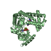
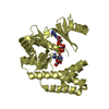
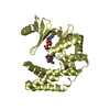
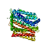
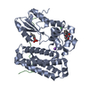
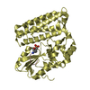
[English] 日本語
Yorodumi
- PDB-3ob2: Crystal structure of c-Cbl TKB domain in complex with double phos... -

+
Open data


ID or keywords:

Loading...

-
Basic information

Entry
Database: PDB / ID: 3ob2
TitleCrystal structure of c-Cbl TKB domain in complex with double phosphorylated EGFR peptide
Components
  • 12-meric peptide from Epidermal growth factor receptor
  • E3 ubiquitin-protein ligase CBL
KeywordsLigase/signaling Protein / protein-peptide complex / double phosphorylation / Ligase-signaling Protein complex
Function / homology
Function and homology information


regulation of platelet-derived growth factor receptor-alpha signaling pathway / ubiquitin-dependent endocytosis / regulation of Rap protein signal transduction / entry of bacterium into host cell / flotillin complex / phosphatidylinositol 3-kinase regulatory subunit binding / response to hydroxyisoflavone / multivesicular body, internal vesicle lumen / positive regulation of prolactin secretion / negative regulation of cardiocyte differentiation ...regulation of platelet-derived growth factor receptor-alpha signaling pathway / ubiquitin-dependent endocytosis / regulation of Rap protein signal transduction / entry of bacterium into host cell / flotillin complex / phosphatidylinositol 3-kinase regulatory subunit binding / response to hydroxyisoflavone / multivesicular body, internal vesicle lumen / positive regulation of prolactin secretion / negative regulation of cardiocyte differentiation / positive regulation of protein kinase C activity / diterpenoid metabolic process / Shc-EGFR complex / ovulation cycle / Inhibition of Signaling by Overexpressed EGFR / epidermal growth factor receptor activity / EGFR interacts with phospholipase C-gamma / positive regulation of mucus secretion / response to UV-A / epidermal growth factor binding / positive regulation of epidermal growth factor receptor signaling pathway / PLCG1 events in ERBB2 signaling / tongue development / midgut development / ERBB2-EGFR signaling pathway / hydrogen peroxide metabolic process / PTK6 promotes HIF1A stabilization / digestive tract morphogenesis / regulation of nitric-oxide synthase activity / Regulation of KIT signaling / ERBB2 Activates PTK6 Signaling / morphogenesis of an epithelial fold / mast cell degranulation / intracellular vesicle / Signaling by EGFR / response to testosterone / Interleukin-6 signaling / response to cobalamin / negative regulation of epidermal growth factor receptor signaling pathway / response to starvation / transmembrane receptor protein tyrosine kinase activator activity / protein tyrosine kinase activator activity / cellular response to platelet-derived growth factor stimulus / Signaling by ERBB4 / regulation of phosphatidylinositol 3-kinase/protein kinase B signal transduction / protein insertion into membrane / eyelid development in camera-type eye / protein monoubiquitination / TGF-beta receptor signaling activates SMADs / cerebral cortex cell migration / ERBB2 Regulates Cell Motility / regulation of JNK cascade / activation of phospholipase C activity / positive regulation of cyclin-dependent protein serine/threonine kinase activity / PI3K events in ERBB2 signaling / negative regulation of mitotic cell cycle / hair follicle development / MAP kinase kinase kinase activity / Estrogen-dependent nuclear events downstream of ESR-membrane signaling / positive regulation of G1/S transition of mitotic cell cycle / embryonic placenta development / positive regulation of bone resorption / GAB1 signalosome / protein autoubiquitination / positive regulation of nitric oxide mediated signal transduction / salivary gland morphogenesis / regulation of peptidyl-tyrosine phosphorylation / peptidyl-tyrosine autophosphorylation / positive regulation of glial cell proliferation / positive regulation of phosphorylation / positive regulation of vasoconstriction / FLT3 signaling by CBL mutants / Signaling by ERBB2 / cellular response to epidermal growth factor stimulus / cellular response to cadmium ion / PTK6 Regulates RTKs and Their Effectors AKT1 and DOK1 / Negative regulation of FLT3 / positive regulation of DNA repair / GRB2 events in EGFR signaling / SHC1 events in EGFR signaling / EGFR Transactivation by Gastrin / TFAP2 (AP-2) family regulates transcription of growth factors and their receptors / GRB2 events in ERBB2 signaling / phosphotyrosine residue binding / transmembrane receptor protein tyrosine kinase activity / neurogenesis / ephrin receptor binding / cellular response to dexamethasone stimulus / SHC1 events in ERBB2 signaling / ossification / positive regulation of synaptic transmission, glutamatergic / basal plasma membrane / cellular response to nerve growth factor stimulus / regulation of ERK1 and ERK2 cascade / InlB-mediated entry of Listeria monocytogenes into host cell / neuron projection morphogenesis / positive regulation of superoxide anion generation / positive regulation of DNA replication / response to activity / Signal transduction by L1
Similarity search - Function
Adaptor protein Cbl, N-terminal domain / Adaptor protein Cbl, N-terminal helical / Adaptor protein Cbl, EF hand-like / Adaptor protein Cbl, SH2-like domain / Adaptor protein Cbl, PTB domain / Adaptor protein Cbl / CBL proto-oncogene N-terminal domain 1 / CBL proto-oncogene N-terminus, EF hand-like domain / CBL proto-oncogene N-terminus, SH2-like domain / Cbl-type phosphotyrosine-binding (Cbl-PTB) domain profile. ...Adaptor protein Cbl, N-terminal domain / Adaptor protein Cbl, N-terminal helical / Adaptor protein Cbl, EF hand-like / Adaptor protein Cbl, SH2-like domain / Adaptor protein Cbl, PTB domain / Adaptor protein Cbl / CBL proto-oncogene N-terminal domain 1 / CBL proto-oncogene N-terminus, EF hand-like domain / CBL proto-oncogene N-terminus, SH2-like domain / Cbl-type phosphotyrosine-binding (Cbl-PTB) domain profile. / Adaptor protein Cbl, N-terminal domain superfamily / Transcription Elongation Factor S-II; Chain A / UBA/TS-N domain / Ubiquitin associated domain / Zinc finger, C3HC4 RING-type / Zinc finger, C3HC4 type (RING finger) / Ubiquitin-associated domain / Ubiquitin-associated domain (UBA) profile. / : / Epidermal growth factor receptor transmembrane-juxtamembrane segment / Tyrosine protein kinase, EGF/ERB/XmrK receptor / Growth factor receptor domain 4 / Growth factor receptor domain IV / UBA-like superfamily / SH2 domain / SHC Adaptor Protein / Receptor L-domain / Furin-like cysteine-rich domain / Receptor L-domain superfamily / Furin-like cysteine rich region / Receptor L domain / Furin-like repeat / Furin-like repeats / EF-hand / Zinc finger, RING-type, conserved site / Zinc finger RING-type signature. / Recoverin; domain 1 / Ring finger / Growth factor receptor cysteine-rich domain superfamily / Zinc finger RING-type profile. / Zinc finger, RING-type / SH2 domain superfamily / EF-hand domain pair / Tyrosine-protein kinase, catalytic domain / Tyrosine kinase, catalytic domain / Tyrosine protein kinases specific active-site signature. / Tyrosine-protein kinase, active site / Protein tyrosine and serine/threonine kinase / Serine-threonine/tyrosine-protein kinase, catalytic domain / Zinc finger, RING/FYVE/PHD-type / Protein kinase, ATP binding site / Protein kinases ATP-binding region signature. / Protein kinase domain profile. / Protein kinase domain / Protein kinase-like domain superfamily / Up-down Bundle / 2-Layer Sandwich / Orthogonal Bundle / Mainly Alpha / Alpha Beta
Similarity search - Domain/homology
Epidermal growth factor receptor / E3 ubiquitin-protein ligase CBL
Similarity search - Component
Biological speciesHomo sapiens (human)
MethodX-RAY DIFFRACTION / MOLECULAR REPLACEMENT / molecular replacement / Resolution: 2.1 Å
AuthorsSun, Q. / Sivaraman, J.
CitationJournal: Plos One / Year: 2010
Title: Additional serine/threonine phosphorylation reduces binding affinity but preserves interface topography of substrate proteins to the c-Cbl TKB domain
Authors: Sun, Q. / Jackson, R.A. / Ng, C. / Guy, G.R. / Sivaraman, J.
History
DepositionAug 6, 2010Deposition site: RCSB / Processing site: PDBJ
Revision 1.0Aug 18, 2010Provider: repository / Type: Initial release
Revision 1.1Jul 13, 2011Group: Version format compliance
Revision 1.2Nov 8, 2017Group: Refinement description / Category: software
Revision 1.3Nov 1, 2023Group: Data collection / Database references ...Data collection / Database references / Derived calculations / Refinement description
Category: chem_comp_atom / chem_comp_bond ...chem_comp_atom / chem_comp_bond / database_2 / pdbx_initial_refinement_model / struct_conn / struct_ref_seq_dif
Item: _database_2.pdbx_DOI / _database_2.pdbx_database_accession ..._database_2.pdbx_DOI / _database_2.pdbx_database_accession / _struct_conn.pdbx_leaving_atom_flag / _struct_ref_seq_dif.details
Revision 1.4Dec 6, 2023Group: Data collection / Category: chem_comp_atom / chem_comp_bond / Item: _chem_comp_atom.atom_id / _chem_comp_bond.atom_id_2

-
Structure visualization

Structure viewerMolecule:
MolmilJmol/JSmol

Downloads & links

-
Assembly

Deposited unit
A: 12-meric peptide from Epidermal growth factor receptor
B: E3 ubiquitin-protein ligase CBL


Theoretical massNumber of molelcules
Total (without water)39,7692
Polymers39,7692
Non-polymers00
Water5,657314
1


  • Idetical with deposited unit
  • defined by author&software
TypeNameSymmetry operationNumber
identity operation1_555x,y,z1
Buried area1280 Å2
ΔGint-5 kcal/mol
Surface area15200 Å2
MethodPISA
Unit cell
Length a, b, c (Å)122.771, 122.771, 55.425
Angle α, β, γ (deg.)90.000, 90.000, 120.000
Int Tables number168
Space group name H-MP6
Components on special symmetry positions
IDModelComponents
11A-303-

HOH

-
Components

#1: Protein/peptide 12-meric peptide from Epidermal growth factor receptor / Receptor tyrosine-protein kinase erbB-1 / Proto-oncogene c-ErbB-1


Mass: 1576.429 Da / Num. of mol.: 1 / Fragment: residues 1063-1074 / Source method: obtained synthetically / Details: synthetic peptide / Source: (synth.) Homo sapiens (human) / References: UniProt: P00533
#2: Protein E3 ubiquitin-protein ligase CBL / Signal transduction protein CBL / Proto-oncogene c-Cbl / Casitas B-lineage lymphoma proto-oncogene ...Signal transduction protein CBL / Proto-oncogene c-Cbl / Casitas B-lineage lymphoma proto-oncogene / RING finger protein 55


Mass: 38192.090 Da / Num. of mol.: 1 / Fragment: c-Cbl TKB domain, residues 25-351
Source method: isolated from a genetically manipulated source
Source: (gene. exp.) Homo sapiens (human) / Plasmid: pGEX-4T-1 / Production host: Escherichia coli (E. coli) / Strain (production host): BL21(DE3)
References: UniProt: P22681, Ligases; Forming carbon-nitrogen bonds; Acid-amino-acid ligases (peptide synthases)
#3: Water ChemComp-HOH / water / Water


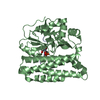
Mass: 18.015 Da / Num. of mol.: 314 / Source method: isolated from a natural source / Formula: H2O

-
Experimental details

-
Experiment

ExperimentMethod: X-RAY DIFFRACTION / Number of used crystals: 1

-
Sample preparation

CrystalDensity Matthews: 3.03 Å3/Da / Density % sol: 59.43 % / Mosaicity: 0.677 °
Crystal growTemperature: 298 K / Method: vapor diffusion, hanging drop / pH: 6.1
Details: 20% (w/v) PEG 3350, 150mM NaK tartrate, 0.1 M Bis-Tris propane pH 6.1, VAPOR DIFFUSION, HANGING DROP, temperature 298K

-
Data collection

DiffractionMean temperature: 93 K
Diffraction sourceSource: ROTATING ANODE / Type: BRUKER PROTEUM X8 / Wavelength: 1.5418 Å
DetectorType: BRUKER AXS MICROSTAR / Detector: CCD / Date: Jun 12, 2009
RadiationProtocol: SINGLE WAVELENGTH / Monochromatic (M) / Laue (L): M / Scattering type: x-ray
Radiation wavelengthWavelength: 1.5418 Å / Relative weight: 1
ReflectionResolution: 2.02→50 Å / Num. obs: 30870 / % possible obs: 98.5 % / Redundancy: 7.2 % / Rmerge(I) obs: 0.062 / Χ2: 1.016 / Net I/σ(I): 20.1
Reflection shell
Resolution (Å)Redundancy (%)Rmerge(I) obsNum. unique allΧ2% possible all
2.02-2.093.20.39929011.06292.7
2.09-2.184.20.34430501.04598.1
2.18-2.274.70.28530541.04698.3
2.27-2.395.30.24431011.02599.3
2.39-2.5460.19130711.00898.8
2.54-2.746.70.14730900.99699.4
2.74-3.027.60.10830970.97799.4
3.02-3.459.40.06931430.98399.6
3.45-4.3511.40.04631381.02199.8
4.35-5012.50.03632251.04399.6

-
Phasing

PhasingMethod: molecular replacement

-
Processing

Software
NameVersionClassificationNB
DENZOdata reduction
SCALEPACKdata scaling
MOLREPphasing
REFMACrefinement
PDB_EXTRACT3.1data extraction
HKL-2000data collection
RefinementMethod to determine structure: MOLECULAR REPLACEMENT
Starting model: PDB ENTRY 3BUM
Resolution: 2.1→19.85 Å / Cor.coef. Fo:Fc: 0.954 / Cor.coef. Fo:Fc free: 0.938 / Occupancy max: 1 / Occupancy min: 0.33 / FOM work R set: 0.8687 / SU B: 4.264 / SU ML: 0.112 / Cross valid method: THROUGHOUT / σ(F): 0 / ESU R: 0.169 / ESU R Free: 0.152 / Stereochemistry target values: MAXIMUM LIKELIHOOD
Details: HYDROGENS HAVE BEEN ADDED IN THE RIDING POSITIONS U VALUES: REFINED INDIVIDUALLY
RfactorNum. reflection% reflectionSelection details
Rfree0.21732 1425 5.1 %RANDOM
Rwork0.18269 ---
obs0.18449 27981 99.81 %-
Solvent computationIon probe radii: 0.8 Å / Shrinkage radii: 0.8 Å / VDW probe radii: 1.4 Å / Solvent model: MASK
Displacement parametersBiso max: 75.15 Å2 / Biso mean: 33.471 Å2 / Biso min: 5.53 Å2
Baniso -1Baniso -2Baniso -3
1--0.86 Å2-0.43 Å20 Å2
2---0.86 Å20 Å2
3---1.29 Å2
Refinement stepCycle: LAST / Resolution: 2.1→19.85 Å
ProteinNucleic acidLigandSolventTotal
Num. atoms2534 0 0 314 2848
Refine LS restraints
Refine-IDTypeDev idealDev ideal targetNumber
X-RAY DIFFRACTIONr_bond_refined_d0.0180.0222602
X-RAY DIFFRACTIONr_bond_other_d
X-RAY DIFFRACTIONr_angle_refined_deg1.6421.9653518
X-RAY DIFFRACTIONr_angle_other_deg
X-RAY DIFFRACTIONr_dihedral_angle_1_deg5.7125305
X-RAY DIFFRACTIONr_dihedral_angle_2_deg36.6123.306124
X-RAY DIFFRACTIONr_dihedral_angle_3_deg17.40615.032465
X-RAY DIFFRACTIONr_dihedral_angle_4_deg21.221518
X-RAY DIFFRACTIONr_chiral_restr0.1060.2373
X-RAY DIFFRACTIONr_gen_planes_refined0.0080.0211966
X-RAY DIFFRACTIONr_gen_planes_other
X-RAY DIFFRACTIONr_nbd_refined
X-RAY DIFFRACTIONr_nbd_other
X-RAY DIFFRACTIONr_nbtor_refined
X-RAY DIFFRACTIONr_nbtor_other
X-RAY DIFFRACTIONr_xyhbond_nbd_refined
X-RAY DIFFRACTIONr_xyhbond_nbd_other
X-RAY DIFFRACTIONr_metal_ion_refined
X-RAY DIFFRACTIONr_metal_ion_other
X-RAY DIFFRACTIONr_symmetry_vdw_refined
X-RAY DIFFRACTIONr_symmetry_vdw_other
X-RAY DIFFRACTIONr_symmetry_hbond_refined
X-RAY DIFFRACTIONr_symmetry_hbond_other
X-RAY DIFFRACTIONr_symmetry_metal_ion_refined
X-RAY DIFFRACTIONr_symmetry_metal_ion_other
X-RAY DIFFRACTIONr_mcbond_it0.9551.51535
X-RAY DIFFRACTIONr_mcbond_other
X-RAY DIFFRACTIONr_mcangle_it1.84222485
X-RAY DIFFRACTIONr_scbond_it2.90431067
X-RAY DIFFRACTIONr_scangle_it4.7144.51033
X-RAY DIFFRACTIONr_rigid_bond_restr
X-RAY DIFFRACTIONr_sphericity_free
X-RAY DIFFRACTIONr_sphericity_bonded
LS refinement shellResolution: 2.1→2.154 Å / Total num. of bins used: 20
RfactorNum. reflection% reflection
Rfree0.309 95 -
Rwork0.268 1937 -
all-2032 -
obs--99.36 %

+
About Yorodumi

-
News

-
Feb 9, 2022. New format data for meta-information of EMDB entries

New format data for meta-information of EMDB entries

  • Version 3 of the EMDB header file is now the official format.
  • The previous official version 1.9 will be removed from the archive.

Related info.:EMDB header

External links:wwPDB to switch to version 3 of the EMDB data model

-
Aug 12, 2020. Covid-19 info

Covid-19 info

URL: https://pdbj.org/emnavi/covid19.php

New page: Covid-19 featured information page in EM Navigator.

Related info.:Covid-19 info / Mar 5, 2020. Novel coronavirus structure data

+
Mar 5, 2020. Novel coronavirus structure data

Novel coronavirus structure data

Related info.:Yorodumi Speices / Aug 12, 2020. Covid-19 info

External links:COVID-19 featured content - PDBj / Molecule of the Month (242):Coronavirus Proteases

+
Jan 31, 2019. EMDB accession codes are about to change! (news from PDBe EMDB page)

EMDB accession codes are about to change! (news from PDBe EMDB page)

  • The allocation of 4 digits for EMDB accession codes will soon come to an end. Whilst these codes will remain in use, new EMDB accession codes will include an additional digit and will expand incrementally as the available range of codes is exhausted. The current 4-digit format prefixed with “EMD-” (i.e. EMD-XXXX) will advance to a 5-digit format (i.e. EMD-XXXXX), and so on. It is currently estimated that the 4-digit codes will be depleted around Spring 2019, at which point the 5-digit format will come into force.
  • The EM Navigator/Yorodumi systems omit the EMD- prefix.

Related info.:Q: What is EMD? / ID/Accession-code notation in Yorodumi/EM Navigator

External links:EMDB Accession Codes are Changing Soon! / Contact to PDBj

+
Jul 12, 2017. Major update of PDB

Major update of PDB

  • wwPDB released updated PDB data conforming to the new PDBx/mmCIF dictionary.
  • This is a major update changing the version number from 4 to 5, and with Remediation, in which all the entries are updated.
  • In this update, many items about electron microscopy experimental information are reorganized (e.g. em_software).
  • Now, EM Navigator and Yorodumi are based on the updated data.

External links:wwPDB Remediation / Enriched Model Files Conforming to OneDep Data Standards Now Available in the PDB FTP Archive

-
Yorodumi

Thousand views of thousand structures

  • Yorodumi is a browser for structure data from EMDB, PDB, SASBDB, etc.
  • This page is also the successor to EM Navigator detail page, and also detail information page/front-end page for Omokage search.
  • The word "yorodu" (or yorozu) is an old Japanese word meaning "ten thousand". "mi" (miru) is to see.

Related info.:EMDB / PDB / SASBDB / Comparison of 3 databanks / Yorodumi Search / Aug 31, 2016. New EM Navigator & Yorodumi / Yorodumi Papers / Jmol/JSmol / Function and homology information / Changes in new EM Navigator and Yorodumi

Read more