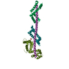
- PDB-3oa7: Structure of the C-terminal domain of Cnm67, a core component of ... -
+
Open data
ID or keywords:
Loading...
-
Basic information
Entry
Database: PDB / ID: 3oa7
Title
Structure of the C-terminal domain of Cnm67, a core component of the spindle pole body of Saccharomyces cerevisiae
Components
Head morphogenesis protein, Chaotic nuclear migration protein 67 fusion protein
Keywords
STRUCTURAL PROTEIN / coiled coils / spindle pole body
Function / homology
Function and homology information
outer plaque of mitotic spindle pole body / spindle pole body organization / outer plaque of spindle pole body / central plaque of spindle pole body / viral scaffold / regulation of microtubule nucleation / spindle pole body / sporulation resulting in formation of a cellular spore / virion assembly / mitotic spindle organization ...outer plaque of mitotic spindle pole body / spindle pole body organization / outer plaque of spindle pole body / central plaque of spindle pole body / viral scaffold / regulation of microtubule nucleation / spindle pole body / sporulation resulting in formation of a cellular spore / virion assembly / mitotic spindle organization / meiotic cell cycle / structural constituent of cytoskeleton / cell division / DNA binding / cytoplasm Similarity search - Function
Septation initiation Sid4-like / Septation initiation / Bacteriophage phi-29 scaffolding protein Gp7 / Capsid assembly scaffolding protein Gp7 / Phi29 scaffolding protein Similarity search - Domain/homology
Method to determine structure: SAD / Resolution: 2.3→20 Å / Cor.coef. Fo:Fc: 0.957 / Cor.coef. Fo:Fc free: 0.947 / SU B: 13.059 / SU ML: 0.133 / Cross valid method: THROUGHOUT / ESU R: 0.265 / ESU R Free: 0.218 / Stereochemistry target values: MAXIMUM LIKELIHOOD / Details: HYDROGENS HAVE BEEN ADDED IN THE RIDING POSITIONS
Rfactor
Num. reflection
% reflection
Selection details
Rfree
0.27069
719
5.1 %
RANDOM
Rwork
0.23765
-
-
-
obs
0.23931
13508
99.66 %
-
all
-
13554
-
-
Solvent computation
Ion probe radii: 0.8 Å / Shrinkage radii: 0.9 Å / VDW probe radii: 1 Å / Solvent model: BABINET MODEL WITH MASK
Displacement parameters
Biso mean: 81.842 Å2
Baniso -1
Baniso -2
Baniso -3
1-
2.43 Å2
0 Å2
0 Å2
2-
-
-1.39 Å2
0 Å2
3-
-
-
-1.04 Å2
Refinement step
Cycle: LAST / Resolution: 2.3→20 Å
Protein
Nucleic acid
Ligand
Solvent
Total
Num. atoms
1585
0
0
18
1603
Refine LS restraints
Refine-ID
Type
Dev ideal
Dev ideal target
Number
X-RAY DIFFRACTION
r_bond_refined_d
0.019
0.022
1610
X-RAY DIFFRACTION
r_angle_refined_deg
1.677
1.977
2170
X-RAY DIFFRACTION
r_dihedral_angle_1_deg
6.058
5
191
X-RAY DIFFRACTION
r_dihedral_angle_2_deg
37.978
24.588
85
X-RAY DIFFRACTION
r_dihedral_angle_3_deg
20.573
15
310
X-RAY DIFFRACTION
r_dihedral_angle_4_deg
16.645
15
13
X-RAY DIFFRACTION
r_chiral_restr
0.114
0.2
244
X-RAY DIFFRACTION
r_gen_planes_refined
0.007
0.021
1207
X-RAY DIFFRACTION
r_mcbond_it
5.283
6
965
X-RAY DIFFRACTION
r_mcangle_it
8.62
60
1565
X-RAY DIFFRACTION
r_scbond_it
9.194
8
645
X-RAY DIFFRACTION
r_scangle_it
14.292
80
605
LS refinement shell
Resolution: 2.3→2.359 Å / Total num. of bins used: 20
Rfactor
Num. reflection
% reflection
Rfree
0.287
51
-
Rwork
0.272
955
-
obs
-
-
99.31 %
Refinement TLS params.
Method: refined / Refine-ID: X-RAY DIFFRACTION
ID
L11 (°2)
L12 (°2)
L13 (°2)
L22 (°2)
L23 (°2)
L33 (°2)
S11 (Å °)
S12 (Å °)
S13 (Å °)
S21 (Å °)
S22 (Å °)
S23 (Å °)
S31 (Å °)
S32 (Å °)
S33 (Å °)
T11 (Å2)
T12 (Å2)
T13 (Å2)
T22 (Å2)
T23 (Å2)
T33 (Å2)
Origin x (Å)
Origin y (Å)
Origin z (Å)
1
2.9674
0.1714
0.7645
13.2604
-3.9445
4.6061
-0.19
-0.61
0.2372
0.3903
0.1918
0.3283
-0.0583
-0.1271
-0.0018
0.1474
0.0493
0.0541
0.2837
-0.022
0.2112
27.094
72.834
16.602
2
1.7156
-0.0161
-1.25
1.5226
0.7765
2.7788
0.2716
-0.0396
-0.0023
0.0051
-0.0431
0.0244
-0.0608
0.0449
-0.2286
0.0555
0.0082
-0.0212
0.2361
0.0099
0.0515
30.394
145.288
15.365
Refinement TLS group
ID
Refine-ID
Refine TLS-ID
Auth asym-ID
Auth seq-ID
1
X-RAY DIFFRACTION
1
A
1 - 50
2
X-RAY DIFFRACTION
1
A
429 - 432
3
X-RAY DIFFRACTION
2
A
433 - 573
+
About Yorodumi
-
News
-
Feb 9, 2022. New format data for meta-information of EMDB entries
New format data for meta-information of EMDB entries
Version 3 of the EMDB header file is now the official format.
The previous official version 1.9 will be removed from the archive.
In the structure databanks used in Yorodumi, some data are registered as the other names, "COVID-19 virus" and "2019-nCoV". Here are the details of the virus and the list of structure data.
Jan 31, 2019. EMDB accession codes are about to change! (news from PDBe EMDB page)
EMDB accession codes are about to change! (news from PDBe EMDB page)
The allocation of 4 digits for EMDB accession codes will soon come to an end. Whilst these codes will remain in use, new EMDB accession codes will include an additional digit and will expand incrementally as the available range of codes is exhausted. The current 4-digit format prefixed with “EMD-” (i.e. EMD-XXXX) will advance to a 5-digit format (i.e. EMD-XXXXX), and so on. It is currently estimated that the 4-digit codes will be depleted around Spring 2019, at which point the 5-digit format will come into force.
The EM Navigator/Yorodumi systems omit the EMD- prefix.
Related info.:Q: What is EMD? / ID/Accession-code notation in Yorodumi/EM Navigator
Yorodumi is a browser for structure data from EMDB, PDB, SASBDB, etc.
This page is also the successor to EM Navigator detail page, and also detail information page/front-end page for Omokage search.
The word "yorodu" (or yorozu) is an old Japanese word meaning "ten thousand". "mi" (miru) is to see.
Related info.:EMDB / PDB / SASBDB / Comparison of 3 databanks / Yorodumi Search / Aug 31, 2016. New EM Navigator & Yorodumi / Yorodumi Papers / Jmol/JSmol / Function and homology information / Changes in new EM Navigator and Yorodumi
 Movie
Movie Controller

 Yorodumi
 Open data
 Basic information
 Components
 Keywords
 Function and homology information
 Bacillus phage phi29 (virus)
 X-RAY DIFFRACTION /
  Authors
 Citation
 Structure visualization
 Downloads & links
 Other downloads


 PDBj
PDBj Assembly

 Mass: 18.015 Da / Num. of mol.: 18 / Source method: isolated from a natural source / Formula: H2O
 Sample preparation
 / Beamline: 19-BM / Wavelength: 0.97921 Å
 Processing