 Movie
Movie Controller

[English] 日本語
 Yorodumi
- PDB-3o98: Glutathionylspermidine synthetase/amidase C59A complex with ADP a... -
+ Open data

- Basic information
| Entry | Database: PDB / ID: 3o98 | ||||||
|---|---|---|---|---|---|---|---|
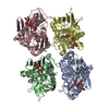
| Title | Glutathionylspermidine synthetase/amidase C59A complex with ADP and Gsp | ||||||
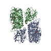
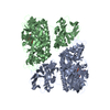
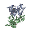
 Components Components | Bifunctional glutathionylspermidine synthetase/amidase | ||||||
 Keywords Keywords | Ligase / Hydrolase | ||||||
| Function / homology |  Function and homology information Function and homology informationglutathionylspermidine amidase / glutathionylspermidine synthase / glutathionylspermidine amidase activity / glutathionylspermidine synthase activity / spermidine metabolic process / glutathione metabolic process / ATP binding / metal ion binding / cytosol Similarity search - Function | ||||||
| Biological species |  | ||||||
| Method |  X-RAY DIFFRACTION / SYNCHROTRON / MOLECULAR REPLACEMENT / Resolution: 2.8 Å X-RAY DIFFRACTION / SYNCHROTRON / MOLECULAR REPLACEMENT / Resolution: 2.8 Å | ||||||
 Authors Authors | Pai, C.H. / Lin, C.H. / Wang, A.H.-J. | ||||||
 Citation Citation | Journal: Protein Sci. / Year: 2011 Title: Structure and mechanism of Escherichia coli glutathionylspermidine amidase belonging to the family of cysteine; histidine-dependent amidohydrolases/peptidases Authors: Pai, C.-H. / Wu, H.-J. / Lin, C.-H. / Wang, A.H.-J. #1: Journal: Embo J. / Year: 2006Title: Dual binding sites for translocation catalysis by Escherichia coli glutathionylspermidine synthetase Authors: Pai, C.H. / Chiang, B.Y. / Ko, T.P. / Chou, C.C. / Chong, C.M. / Yen, F.J. / Chen, S. / Coward, J.K. / Wang, A.H.-J. / Lin, C.H. | ||||||
| History |
|
- Structure visualization
| Structure viewer | Molecule: MolmilJmol/JSmol |
|---|
- Downloads & links
-Download
| PDBx/mmCIF format | 3o98.cif.gz | 258.5 KB | Display | PDBx/mmCIF format |
|---|---|---|---|---|
| PDB format | pdb3o98.ent.gz | 204.5 KB | Display | PDB format |
| PDBx/mmJSON format | 3o98.json.gz | Tree view | PDBx/mmJSON format | |
| Others |  Other downloads Other downloads |
-Validation report
| Arichive directory | https://data.pdbj.org/pub/pdb/validation_reports/o9/3o98ftp://data.pdbj.org/pub/pdb/validation_reports/o9/3o98 | HTTPS FTP |
|---|
-Related structure data
| Related structure data |  3a2yC 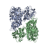 2iobS S: Starting model for refinement C: citing same article ( |
|---|---|
| Similar structure data |


-Links
 PDBj
PDBj


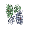
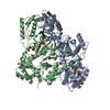
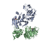
- Assembly
| Deposited unit | 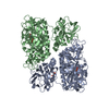
| ||||||||
|---|---|---|---|---|---|---|---|---|---|
| 1 |
| ||||||||
| Unit cell |
|
-Components
| #1: Protein | Mass: 70579.672 Da / Num. of mol.: 2 / Mutation: C59A Source method: isolated from a genetically manipulated source Source: (gene. exp.) References: UniProt: P0AES0, glutathionylspermidine synthase, glutathionylspermidine amidase #2: Chemical | ChemComp-TS5 / | 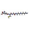  Mass: 434.554 Da / Num. of mol.: 1 / Source method: obtained synthetically / Formula: C17H34N6O5S Mass: 434.554 Da / Num. of mol.: 1 / Source method: obtained synthetically / Formula: C17H34N6O5S#3: Chemical |   Mass: 427.201 Da / Num. of mol.: 2 / Source method: obtained synthetically / Formula: C10H15N5O10P2 / Comment: ADP, energy-carrying molecule*YM Mass: 427.201 Da / Num. of mol.: 2 / Source method: obtained synthetically / Formula: C10H15N5O10P2 / Comment: ADP, energy-carrying molecule*YM#4: Chemical | ChemComp-MG /   Mass: 24.305 Da / Num. of mol.: 4 / Source method: obtained synthetically / Formula: Mg Mass: 24.305 Da / Num. of mol.: 4 / Source method: obtained synthetically / Formula: Mg#5: Water | ChemComp-HOH / |  Mass: 18.015 Da / Num. of mol.: 206 / Source method: isolated from a natural source / Formula: H2O Mass: 18.015 Da / Num. of mol.: 206 / Source method: isolated from a natural source / Formula: H2O |
|---|
-Experimental details
-Experiment
| Experiment | Method: X-RAY DIFFRACTION / Number of used crystals: 1 |
|---|
- Sample preparation
| Crystal | Density Matthews: 2.48 Å3/Da / Density % sol: 50.38 % |
|---|---|
| Crystal grow | Temperature: 298 K / Method: vapor diffusion, hanging drop / pH: 8.5 Details: 12% PEG 3350, 0.5M MgCl2, 0.1M Tris pH8.5, VAPOR DIFFUSION, HANGING DROP, temperature 298K |
-Data collection
| Diffraction | Mean temperature: 100 K |
|---|---|
| Diffraction source | Source: SYNCHROTRON / Site: NSRRC  / Beamline: BL13B1 / Wavelength: 1 Å / Beamline: BL13B1 / Wavelength: 1 Å |
| Detector | Type: ADSC QUANTUM 315 / Detector: CCD / Date: Aug 31, 2007 |
| Radiation | Protocol: SINGLE WAVELENGTH / Monochromatic (M) / Laue (L): M / Scattering type: x-ray |
| Radiation wavelength | Wavelength: 1 Å / Relative weight: 1 |
| Reflection | Resolution: 2.8→30 Å / Num. all: 32519 / Num. obs: 31316 / % possible obs: 96.3 % / Observed criterion σ(F): 0 / Observed criterion σ(I): 1 / Redundancy: 3.9 % / Rmerge(I) obs: 0.096 / Net I/σ(I): 15.9 |
| Reflection shell | Resolution: 2.8→2.9 Å / % possible all: 94.7 |
- Processing
| Software |
| ||||||||||||||||||||
|---|---|---|---|---|---|---|---|---|---|---|---|---|---|---|---|---|---|---|---|---|---|
| Refinement | Method to determine structure: MOLECULAR REPLACEMENT Starting model: PDB ENTRY 2iob Resolution: 2.8→30 Å / Cross valid method: THROUGHOUT / σ(F): 2 / Stereochemistry target values: Engh & Huber
| ||||||||||||||||||||
| Refine analyze |
| ||||||||||||||||||||
| Refinement step | Cycle: LAST / Resolution: 2.8→30 Å
| ||||||||||||||||||||
| Refine LS restraints |
| ||||||||||||||||||||
| LS refinement shell | Resolution: 2.8→2.9 Å / Rfactor Rfree: 0.3278 / Rfactor Rwork: 0.2808 |
