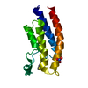
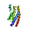
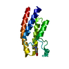
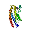
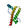
- PDB-3o81: Beta2-microglobulin from Gallus gallus -
+
Open data
ID or keywords:
Loading...
-
Basic information
Entry
Database: PDB / ID: 3o81
Title
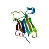
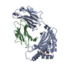
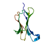
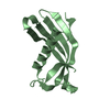
Beta2-microglobulin from Gallus gallus
Components
Beta-2-microglobulin
Keywords
IMMUNE SYSTEM / Ig-like C1-type (immunoglobulin-like) domain / HISTOCOMPATIBILITY ANTIGEN / MHC class I molecule from B21 chicken
Function / homology
Function and homology information
ER-Phagosome pathway / Endosomal/Vacuolar pathway / Immunoregulatory interactions between a Lymphoid and a non-Lymphoid cell / Antigen Presentation: Folding, assembly and peptide loading of class I MHC / antigen processing and presentation of exogenous peptide antigen via MHC class Ib / Neutrophil degranulation / negative regulation of iron ion transport / negative regulation of forebrain neuron differentiation / peptide antigen assembly with MHC class I protein complex / HFE-transferrin receptor complex ...ER-Phagosome pathway / Endosomal/Vacuolar pathway / Immunoregulatory interactions between a Lymphoid and a non-Lymphoid cell / Antigen Presentation: Folding, assembly and peptide loading of class I MHC / antigen processing and presentation of exogenous peptide antigen via MHC class Ib / Neutrophil degranulation / negative regulation of iron ion transport / negative regulation of forebrain neuron differentiation / peptide antigen assembly with MHC class I protein complex / HFE-transferrin receptor complex / MHC class I peptide loading complex / transferrin transport / negative regulation of receptor-mediated endocytosis / cellular response to iron ion / positive regulation of T cell cytokine production / antigen processing and presentation of endogenous peptide antigen via MHC class I / MHC class I protein complex / peptide antigen assembly with MHC class II protein complex / negative regulation of neurogenesis / cellular response to nicotine / MHC class II protein complex / positive regulation of receptor-mediated endocytosis / negative regulation of epithelial cell proliferation / antigen processing and presentation of exogenous peptide antigen via MHC class II / positive regulation of immune response / peptide antigen binding / positive regulation of T cell activation / positive regulation of cellular senescence / MHC class II protein complex binding / late endosome membrane / amyloid fibril formation / protein homotetramerization / intracellular iron ion homeostasis / learning or memory / immune response / lysosomal membrane / structural molecule activity / Golgi apparatus / protein homodimerization activity / extracellular region Similarity search - Function
Resolution: 2→36.85 Å / Cor.coef. Fo:Fc: 0.94 / Cor.coef. Fo:Fc free: 0.913 / SU B: 7.825 / SU ML: 0.12 / Cross valid method: THROUGHOUT / ESU R Free: 0.184 / Stereochemistry target values: MAXIMUM LIKELIHOOD / Details: HYDROGENS HAVE BEEN ADDED IN THE RIDING POSITIONS
Rfactor
Num. reflection
% reflection
Selection details
Rfree
0.24189
667
5 %
RANDOM
Rwork
0.19493
-
-
-
obs
0.19723
12672
100 %
-
all
-
13485
-
-
Solvent computation
Ion probe radii: 0.8 Å / Shrinkage radii: 0.8 Å / VDW probe radii: 1.4 Å / Solvent model: MASK
Displacement parameters
Biso mean: 18.99 Å2
Baniso -1
Baniso -2
Baniso -3
1-
0.44 Å2
0 Å2
1.37 Å2
2-
-
-0.7 Å2
0 Å2
3-
-
-
0.43 Å2
Refinement step
Cycle: LAST / Resolution: 2→36.85 Å
Protein
Nucleic acid
Ligand
Solvent
Total
Num. atoms
1526
0
0
160
1686
Refine LS restraints
Refine-ID
Type
Dev ideal
Dev ideal target
Number
X-RAY DIFFRACTION
r_bond_refined_d
0.011
0.022
1674
X-RAY DIFFRACTION
r_angle_refined_deg
1.271
1.937
2304
X-RAY DIFFRACTION
r_dihedral_angle_1_deg
6.251
5
222
X-RAY DIFFRACTION
r_dihedral_angle_2_deg
37.579
24.667
75
X-RAY DIFFRACTION
r_dihedral_angle_3_deg
11.948
15
269
X-RAY DIFFRACTION
r_dihedral_angle_4_deg
7.1
15
4
X-RAY DIFFRACTION
r_chiral_restr
0.089
0.2
243
X-RAY DIFFRACTION
r_gen_planes_refined
0.007
0.021
1312
X-RAY DIFFRACTION
r_mcbond_it
0.577
1.5
1019
X-RAY DIFFRACTION
r_mcangle_it
1.087
2
1672
X-RAY DIFFRACTION
r_scbond_it
1.616
3
655
X-RAY DIFFRACTION
r_scangle_it
2.564
4.5
617
LS refinement shell
Resolution: 2→2.052 Å / Total num. of bins used: 20
Rfactor
Num. reflection
% reflection
Rfree
0.219
48
-
Rwork
0.193
924
-
obs
-
-
100 %
Refinement TLS params.
Method: refined / Refine-ID: X-RAY DIFFRACTION
ID
L11 (°2)
L12 (°2)
L13 (°2)
L22 (°2)
L23 (°2)
L33 (°2)
S11 (Å °)
S12 (Å °)
S13 (Å °)
S21 (Å °)
S22 (Å °)
S23 (Å °)
S31 (Å °)
S32 (Å °)
S33 (Å °)
T11 (Å2)
T12 (Å2)
T13 (Å2)
T22 (Å2)
T23 (Å2)
T33 (Å2)
Origin x (Å)
Origin y (Å)
Origin z (Å)
1
2.4392
0.0873
-1.2629
2.0147
-0.0129
2.6452
-0.0194
0.0543
-0.0135
0.0571
0.0379
0.1343
-0.0229
-0.1715
-0.0184
0.0033
0.0019
-0.0006
0.0286
0.0018
0.0419
9.7627
-7.6259
2.4487
2
2.0555
-0.2647
1.332
2.6793
0.2427
3.5443
-0.0321
-0.0794
0.0523
-0.0962
0.061
0.137
0.0367
-0.2669
-0.0289
0.0148
-0.0091
0.0075
0.0442
0.0053
0.043
7.0491
0.0323
41.1402
Refinement TLS group
ID
Refine-ID
Refine TLS-ID
Auth asym-ID
Auth seq-ID
1
X-RAY DIFFRACTION
1
A
1 - 97
2
X-RAY DIFFRACTION
2
B
1 - 97
+
About Yorodumi
-
News
-
Feb 9, 2022. New format data for meta-information of EMDB entries
New format data for meta-information of EMDB entries
Version 3 of the EMDB header file is now the official format.
The previous official version 1.9 will be removed from the archive.
In the structure databanks used in Yorodumi, some data are registered as the other names, "COVID-19 virus" and "2019-nCoV". Here are the details of the virus and the list of structure data.
Jan 31, 2019. EMDB accession codes are about to change! (news from PDBe EMDB page)
EMDB accession codes are about to change! (news from PDBe EMDB page)
The allocation of 4 digits for EMDB accession codes will soon come to an end. Whilst these codes will remain in use, new EMDB accession codes will include an additional digit and will expand incrementally as the available range of codes is exhausted. The current 4-digit format prefixed with “EMD-” (i.e. EMD-XXXX) will advance to a 5-digit format (i.e. EMD-XXXXX), and so on. It is currently estimated that the 4-digit codes will be depleted around Spring 2019, at which point the 5-digit format will come into force.
The EM Navigator/Yorodumi systems omit the EMD- prefix.
Related info.:Q: What is EMD? / ID/Accession-code notation in Yorodumi/EM Navigator
Yorodumi is a browser for structure data from EMDB, PDB, SASBDB, etc.
This page is also the successor to EM Navigator detail page, and also detail information page/front-end page for Omokage search.
The word "yorodu" (or yorozu) is an old Japanese word meaning "ten thousand". "mi" (miru) is to see.
Related info.:EMDB / PDB / SASBDB / Comparison of 3 databanks / Yorodumi Search / Aug 31, 2016. New EM Navigator & Yorodumi / Yorodumi Papers / Jmol/JSmol / Function and homology information / Changes in new EM Navigator and Yorodumi
 Movie
Movie Controller


 Open data
 Basic information
 Components
 Keywords
 Function and homology information
 X-RAY DIFFRACTION /
  Authors
 Citation
 Structure visualization
 Downloads & links
 Other downloads


 PDBj
PDBj Assembly


 Mass: 18.015 Da / Num. of mol.: 160 / Source method: isolated from a natural source / Formula: H2O
 Sample preparation
 / Beamline: 14.2 / Wavelength: 0.9184 Å
 Processing