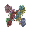
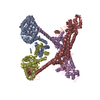
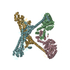
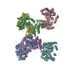
Entry Database : PDB / ID : 3o4xTitle Crystal structure of complex between amino and carboxy terminal fragments of mDia1 (Protein diaphanous homolog 1) x 2 Keywords / / / Function / homology Function Domain/homology Component
/ / / / / / / / / / / / / / / / / / / / / / / / / / / / / / / / / / / / / / / / / / / / / / / / / / / / / / / / / / / / / / / / / / / / / / / / / / / / / / / / / / / / / / / / / / / / / / / / / Biological species Mus musculus (house mouse)Method / / / Resolution : 3.2 Å Authors Eck, M.J. / Nezami, A. / Toms, A.V. Journal : Plos One / Year : 2010Title : Crystal structure of a complex between amino and carboxy terminal fragments of mDia1: insights into autoinhibition of diaphanous-related formins.Authors : Nezami, A. / Poy, F. / Toms, A. / Zheng, W. / Eck, M.J. History Deposition Jul 27, 2010 Deposition site / Processing site Revision 1.0 Oct 13, 2010 Provider / Type Revision 1.1 Jul 13, 2011 Group Revision 1.2 Aug 17, 2011 Group Revision 1.3 Oct 5, 2011 Group Revision 1.4 Apr 11, 2012 Group Revision 1.5 Nov 6, 2024 Group Data collection / Database references ... Data collection / Database references / Refinement description / Structure summary Category chem_comp_atom / chem_comp_bond ... chem_comp_atom / chem_comp_bond / database_2 / pdbx_entry_details / pdbx_modification_feature / struct_ncs_dom_lim / struct_ref_seq_dif Item _database_2.pdbx_DOI / _database_2.pdbx_database_accession ... _database_2.pdbx_DOI / _database_2.pdbx_database_accession / _struct_ncs_dom_lim.beg_auth_comp_id / _struct_ncs_dom_lim.beg_label_asym_id / _struct_ncs_dom_lim.beg_label_comp_id / _struct_ncs_dom_lim.beg_label_seq_id / _struct_ncs_dom_lim.end_auth_comp_id / _struct_ncs_dom_lim.end_label_asym_id / _struct_ncs_dom_lim.end_label_comp_id / _struct_ncs_dom_lim.end_label_seq_id / _struct_ref_seq_dif.details
Show all Show less
 Movie
Movie Controller

 Yorodumi
 Open data
 Basic information
 Components
 Keywords
 Function and homology information
 X-RAY DIFFRACTION /
  Authors
 Citation
 Structure visualization
 Downloads & links
 Other downloads
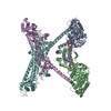


 PDBj
PDBj


 Assembly