 Movie
Movie Controller


+ Open data

- Basic information
| Entry | Database: PDB / ID: 3o0p | ||||||
|---|---|---|---|---|---|---|---|
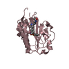
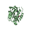
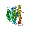
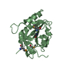
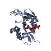
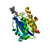
| Title | Pilus-related Sortase C of Group B Streptococcus | ||||||
 Components Components | Sortase family protein | ||||||
 Keywords Keywords | TRANSFERASE / HYDROLASE / sortase / pili assembly / pili subunits | ||||||
| Function / homology |  Function and homology information Function and homology information | ||||||
| Biological species |  Streptococcus agalactiae (bacteria) Streptococcus agalactiae (bacteria) | ||||||
| Method |  X-RAY DIFFRACTION / SYNCHROTRON / MOLECULAR REPLACEMENT / Resolution: 1.3 Å X-RAY DIFFRACTION / SYNCHROTRON / MOLECULAR REPLACEMENT / Resolution: 1.3 Å | ||||||
 Authors Authors | Malito, E. / Spraggon, G. | ||||||
 Citation Citation | Journal: Faseb J. / Year: 2011 Title: Structure analysis and site-directed mutagenesis of defined key residues and motives for pilus-related sortase C1 in group B Streptococcus. Authors: Cozzi, R. / Malito, E. / Nuccitelli, A. / D'Onofrio, M. / Martinelli, M. / Ferlenghi, I. / Grandi, G. / Telford, J.L. / Maione, D. / Rinaudo, C.D. | ||||||
| History |
|
- Structure visualization
| Structure viewer | Molecule: MolmilJmol/JSmol |
|---|
- Downloads & links
-Download
| PDBx/mmCIF format | 3o0p.cif.gz | 89.6 KB | Display | PDBx/mmCIF format |
|---|---|---|---|---|
| PDB format | pdb3o0p.ent.gz | 67.2 KB | Display | PDB format |
| PDBx/mmJSON format | 3o0p.json.gz | Tree view | PDBx/mmJSON format | |
| Others |  Other downloads Other downloads |
-Validation report
| Arichive directory | https://data.pdbj.org/pub/pdb/validation_reports/o0/3o0pftp://data.pdbj.org/pub/pdb/validation_reports/o0/3o0p | HTTPS FTP |
|---|
-Related structure data
| Related structure data |  2w1jS S: Starting model for refinement |
|---|---|
| Similar structure data |


-Links
 PDBj
PDBj


- Assembly
| Deposited unit | 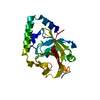
| ||||||||
|---|---|---|---|---|---|---|---|---|---|
| 1 |
| ||||||||
| Unit cell |
|
-Components
| #1: Protein | Mass: 24083.576 Da / Num. of mol.: 1 Source method: isolated from a genetically manipulated source Source: (gene. exp.) Streptococcus agalactiae (bacteria) / Strain: strain 515 (serotype Ia) / Gene: Pilus Island 2a (PI-2a), SAL_1484 / Plasmid: pET 54 DEST / Production host: |
|---|---|
| #2: Water | ChemComp-HOH /  Mass: 18.015 Da / Num. of mol.: 173 / Source method: isolated from a natural source / Formula: H2O Mass: 18.015 Da / Num. of mol.: 173 / Source method: isolated from a natural source / Formula: H2O |
-Experimental details
-Experiment
| Experiment | Method: X-RAY DIFFRACTION / Number of used crystals: 1 |
|---|
- Sample preparation
| Crystal | Density Matthews: 1.78 Å3/Da / Density % sol: 30.88 % |
|---|---|
| Crystal grow | Temperature: 293 K / Method: vapor diffusion, sitting drop / pH: 8.5 Details: 100mM Tris, 200mM NaOAc, 30% PEG-4000, pH 8.5, VAPOR DIFFUSION, SITTING DROP, temperature 293K |
-Data collection
| Diffraction | Mean temperature: 100 K |
|---|---|
| Diffraction source | Source: SYNCHROTRON / Site: ALS  / Beamline: 5.0.3 / Wavelength: 0.9765 Å / Beamline: 5.0.3 / Wavelength: 0.9765 Å |
| Detector | Type: ADSC QUANTUM 315 / Detector: CCD / Date: Oct 27, 2009 |
| Radiation | Monochromator: GRAPHITE / Protocol: SINGLE WAVELENGTH / Monochromatic (M) / Laue (L): M / Scattering type: x-ray |
| Radiation wavelength | Wavelength: 0.9765 Å / Relative weight: 1 |
| Reflection | Resolution: 1.3→50 Å / Num. obs: 40390 / % possible obs: 95 % / Observed criterion σ(F): 2 / Observed criterion σ(I): 2 / Biso Wilson estimate: 11.55 Å2 / Rsym value: 0.09 / Net I/σ(I): 38.2 |
| Reflection shell | Resolution: 1.3→1.35 Å / Redundancy: 4.4 % / Mean I/σ(I) obs: 3 / Rsym value: 0.412 / % possible all: 71.1 |
- Processing
| Software |
| |||||||||||||||||||||||||||||||||||||||||||||||||||||||||||||||||||||||||||||||||||||||||||||||||||||||||
|---|---|---|---|---|---|---|---|---|---|---|---|---|---|---|---|---|---|---|---|---|---|---|---|---|---|---|---|---|---|---|---|---|---|---|---|---|---|---|---|---|---|---|---|---|---|---|---|---|---|---|---|---|---|---|---|---|---|---|---|---|---|---|---|---|---|---|---|---|---|---|---|---|---|---|---|---|---|---|---|---|---|---|---|---|---|---|---|---|---|---|---|---|---|---|---|---|---|---|---|---|---|---|---|---|---|---|
| Refinement | Method to determine structure: MOLECULAR REPLACEMENT Starting model: PDB ENTRY 2W1J Resolution: 1.3→44.781 Å / SU ML: 0.09 / σ(F): 0.18 / Phase error: 16.97 / Stereochemistry target values: ML
| |||||||||||||||||||||||||||||||||||||||||||||||||||||||||||||||||||||||||||||||||||||||||||||||||||||||||
| Solvent computation | Shrinkage radii: 0.9 Å / VDW probe radii: 1.11 Å / Solvent model: FLAT BULK SOLVENT MODEL / Bsol: 59.71 Å2 / ksol: 0.4 e/Å3 | |||||||||||||||||||||||||||||||||||||||||||||||||||||||||||||||||||||||||||||||||||||||||||||||||||||||||
| Displacement parameters |
| |||||||||||||||||||||||||||||||||||||||||||||||||||||||||||||||||||||||||||||||||||||||||||||||||||||||||
| Refinement step | Cycle: LAST / Resolution: 1.3→44.781 Å
| |||||||||||||||||||||||||||||||||||||||||||||||||||||||||||||||||||||||||||||||||||||||||||||||||||||||||
| Refine LS restraints |
| |||||||||||||||||||||||||||||||||||||||||||||||||||||||||||||||||||||||||||||||||||||||||||||||||||||||||
| LS refinement shell |
|
