 Movie
Movie Controller

[English] 日本語
 Yorodumi
- PDB-3nzx: Crystal structure of the yeast 20S proteasome in complex with lig... -
+ Open data

- Basic information
| Entry | Database: PDB / ID: 3nzx | ||||||
|---|---|---|---|---|---|---|---|
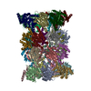
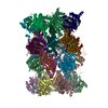
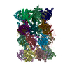
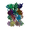
| Title | Crystal structure of the yeast 20S proteasome in complex with ligand 2c | ||||||
 Components Components |
| ||||||
 Keywords Keywords | hydrolase/hydrolase inhibitor / ubiquitin / protein degradation / N-terminal nucleophilic hydrolase / 19S regulatory particle / ubiquitin-tagged substrates / cytosol / nucleus / hydrolase-hydrolase inhibitor complex | ||||||
| Function / homology |  Function and homology information Function and homology informationproteasome core complex assembly / nuclear outer membrane-endoplasmic reticulum membrane network / Proteasome assembly / Cross-presentation of soluble exogenous antigens (endosomes) / TNFR2 non-canonical NF-kB pathway / Ubiquitin-Mediated Degradation of Phosphorylated Cdc25A / proteasomal ubiquitin-independent protein catabolic process / Regulation of PTEN stability and activity / CDK-mediated phosphorylation and removal of Cdc6 / FBXL7 down-regulates AURKA during mitotic entry and in early mitosis ...proteasome core complex assembly / nuclear outer membrane-endoplasmic reticulum membrane network / Proteasome assembly / Cross-presentation of soluble exogenous antigens (endosomes) / TNFR2 non-canonical NF-kB pathway / Ubiquitin-Mediated Degradation of Phosphorylated Cdc25A / proteasomal ubiquitin-independent protein catabolic process / Regulation of PTEN stability and activity / CDK-mediated phosphorylation and removal of Cdc6 / FBXL7 down-regulates AURKA during mitotic entry and in early mitosis / KEAP1-NFE2L2 pathway / Neddylation / Orc1 removal from chromatin / MAPK6/MAPK4 signaling / proteasome storage granule / Antigen processing: Ubiquitination & Proteasome degradation / proteasome endopeptidase complex / endopeptidase activator activity / proteasome core complex, beta-subunit complex / Ub-specific processing proteases / proteasome assembly / threonine-type endopeptidase activity / proteasome core complex, alpha-subunit complex / Neutrophil degranulation / proteasome complex / peroxisome / endopeptidase activity / proteasome-mediated ubiquitin-dependent protein catabolic process / mRNA binding / endoplasmic reticulum membrane / mitochondrion / nucleus / cytosol Similarity search - Function | ||||||
| Biological species |  | ||||||
| Method |  X-RAY DIFFRACTION / SYNCHROTRON / MOLECULAR REPLACEMENT / Resolution: 2.7 Å X-RAY DIFFRACTION / SYNCHROTRON / MOLECULAR REPLACEMENT / Resolution: 2.7 Å | ||||||
 Authors Authors | Groll, M. / Gallastegui, N. / Marechal, X. / Le Ravalec, V. / Basse, N. / Richy, N. / Genin, E. / Huber, R. / Moroder, M. / Vidal, V. / Reboud-Ravaux, M. | ||||||
 Citation Citation | Journal: Chemmedchem / Year: 2010 Title: 20S proteasome inhibition: designing noncovalent linear peptide mimics of the natural product TMC-95A. Authors: Groll, M. / Gallastegui, N. / Marechal, X. / Le Ravalec, V. / Basse, N. / Richy, N. / Genin, E. / Huber, R. / Moroder, L. / Vidal, J. / Reboud-Ravaux, M. | ||||||
| History |
|
- Structure visualization
| Structure viewer | Molecule: MolmilJmol/JSmol |
|---|
- Downloads & links
-Download
| PDBx/mmCIF format | 3nzx.cif.gz | 1.2 MB | Display | PDBx/mmCIF format |
|---|---|---|---|---|
| PDB format | pdb3nzx.ent.gz | 1017 KB | Display | PDB format |
| PDBx/mmJSON format | 3nzx.json.gz | Tree view | PDBx/mmJSON format | |
| Others |  Other downloads Other downloads |
-Validation report
| Summary document | 3nzx_validation.pdf.gz | 688.3 KB | Display | wwPDB validaton report |
|---|---|---|---|---|
| Full document | 3nzx_full_validation.pdf.gz | 883.7 KB | Display | |
| Data in XML | 3nzx_validation.xml.gz | 243.1 KB | Display | |
| Data in CIF | 3nzx_validation.cif.gz | 331.9 KB | Display | |
| Arichive directory | https://data.pdbj.org/pub/pdb/validation_reports/nz/3nzxftp://data.pdbj.org/pub/pdb/validation_reports/nz/3nzx | HTTPS FTP |
-Related structure data
| Related structure data |  3nzjC  3nzwC  1rypS S: Starting model for refinement C: citing same article ( |
|---|---|
| Similar structure data |


-Links
 PDBj
PDBj


- Assembly
| Deposited unit | 
| ||||||||
|---|---|---|---|---|---|---|---|---|---|
| 1 |
| ||||||||
| Unit cell |
| ||||||||
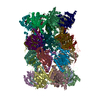
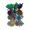
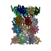
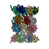
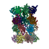
| Details | The asymmetric unit contains the 20S proteasome composed of 28 subunits with 7 different alpha- and 7 different beta-type subunits |
-Components
-Proteasome component ... , 14 types, 28 molecules AOBPCQDRESFTGUHVIWJXKYLZM1N2
| #1: Protein | Mass: 27191.828 Da / Num. of mol.: 2 / Source method: isolated from a natural source / Source: (natural) References: UniProt: P23639, proteasome endopeptidase complex #2: Protein | Mass: 28748.230 Da / Num. of mol.: 2 / Source method: isolated from a natural source / Source: (natural) References: UniProt: P23638, proteasome endopeptidase complex #3: Protein | Mass: 28478.111 Da / Num. of mol.: 2 / Source method: isolated from a natural source / Source: (natural) References: UniProt: P40303, proteasome endopeptidase complex #4: Protein | Mass: 28649.086 Da / Num. of mol.: 2 / Source method: isolated from a natural source / Source: (natural) References: UniProt: P32379, proteasome endopeptidase complex #5: Protein | Mass: 25634.000 Da / Num. of mol.: 2 / Source method: isolated from a natural source / Source: (natural) References: UniProt: P40302, proteasome endopeptidase complex #6: Protein | Mass: 31575.068 Da / Num. of mol.: 2 / Source method: isolated from a natural source / Source: (natural) References: UniProt: P21242, proteasome endopeptidase complex #7: Protein | Mass: 28033.830 Da / Num. of mol.: 2 / Source method: isolated from a natural source / Source: (natural) References: UniProt: P21243, proteasome endopeptidase complex #8: Protein | Mass: 28299.889 Da / Num. of mol.: 2 / Source method: isolated from a natural source / Source: (natural) References: UniProt: P25043, proteasome endopeptidase complex #9: Protein | Mass: 22627.842 Da / Num. of mol.: 2 / Source method: isolated from a natural source / Source: (natural) References: UniProt: P25451, proteasome endopeptidase complex #10: Protein | Mass: 22545.676 Da / Num. of mol.: 2 / Source method: isolated from a natural source / Source: (natural) References: UniProt: P22141, proteasome endopeptidase complex #11: Protein | Mass: 31670.539 Da / Num. of mol.: 2 / Source method: isolated from a natural source / Source: (natural) References: UniProt: P30656, proteasome endopeptidase complex #12: Protein | Mass: 26905.076 Da / Num. of mol.: 2 / Source method: isolated from a natural source / Source: (natural) References: UniProt: P23724, proteasome endopeptidase complex #13: Protein | Mass: 29471.289 Da / Num. of mol.: 2 / Source method: isolated from a natural source / Source: (natural) References: UniProt: P30657, proteasome endopeptidase complex #14: Protein | Mass: 23573.604 Da / Num. of mol.: 2 / Source method: isolated from a natural source / Source: (natural) References: UniProt: P38624, proteasome endopeptidase complex |
|---|
-Protein/peptide / Non-polymers , 2 types, 1336 molecules 34
| #15: Protein/peptide | 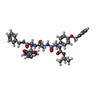  Type: Cyclic peptide / Class: Inhibitor / Mass: 749.851 Da / Num. of mol.: 2 / Source method: obtained synthetically / References: PDB-3NZJ, TMC-95A mimic ligand 2c Type: Cyclic peptide / Class: Inhibitor / Mass: 749.851 Da / Num. of mol.: 2 / Source method: obtained synthetically / References: PDB-3NZJ, TMC-95A mimic ligand 2c#16: Water | ChemComp-HOH / | Mass: 18.015 Da / Num. of mol.: 1334 / Source method: isolated from a natural source / Formula: H2O |
|---|
-Details
| Compound details | THE ENTIRE CHAIN OF 3 OR 4 IS THE POLYMER REPRESENTATION OF THE INHIBITOR COMPOUND UNDER THE ...THE ENTIRE CHAIN OF 3 OR 4 IS THE POLYMER REPRESENTA |
|---|
-Experimental details
-Experiment
| Experiment | Method: X-RAY DIFFRACTION / Number of used crystals: 1 |
|---|
- Sample preparation
| Crystal | Density Matthews: 3.48 Å3/Da / Density % sol: 64.61 % |
|---|---|
| Crystal grow | Temperature: 293 K / Method: vapor diffusion, hanging drop / pH: 6.8 Details: 100 mM MES/Hcl, pH 6.8, 20 mM MGAc2, 12 % MPD, VAPOR DIFFUSION, HANGING DROP, temperature 293K |
-Data collection
| Diffraction | Mean temperature: 100 K |
|---|---|
| Diffraction source | Source: SYNCHROTRON / Site: SLS  / Beamline: X06SA / Wavelength: 1 Å / Beamline: X06SA / Wavelength: 1 Å |
| Detector | Type: MAR CCD 165 mm / Detector: CCD / Date: Nov 4, 2009 |
| Radiation | Monochromator: Focusing Spherical Grating Monochromator / Protocol: SINGLE WAVELENGTH / Monochromatic (M) / Laue (L): M / Scattering type: x-ray |
| Radiation wavelength | Wavelength: 1 Å / Relative weight: 1 |
| Reflection | Resolution: 2.5→50 Å / Num. all: 360910 / Num. obs: 359106 / % possible obs: 99.6 % / Observed criterion σ(F): 2.5 / Observed criterion σ(I): 2.5 / Redundancy: 2.6 % / Biso Wilson estimate: 30.3 Å2 / Rmerge(I) obs: 0.085 / Net I/σ(I): 10.1 |
| Reflection shell | Resolution: 2.5→2.6 Å / Redundancy: 2.4 % / Rmerge(I) obs: 0.533 / Mean I/σ(I) obs: 2.5 / % possible all: 99.7 |
- Processing
| Software |
| |||||||||||||||||||||||||
|---|---|---|---|---|---|---|---|---|---|---|---|---|---|---|---|---|---|---|---|---|---|---|---|---|---|---|
| Refinement | Method to determine structure: MOLECULAR REPLACEMENT Starting model: PDB entry 1RYP Resolution: 2.7→15 Å / Rfactor Rfree error: 0.002 / Data cutoff high absF: 6335150.1 / Data cutoff low absF: 0 / Isotropic thermal model: RESTRAINED / Cross valid method: THROUGHOUT / σ(F): 0 / σ(I): 2.5 / Stereochemistry target values: Engh & Huber / Details: BULK SOLVENT MODEL USED
| |||||||||||||||||||||||||
| Solvent computation | Solvent model: FLAT MODEL / Bsol: 38.9587 Å2 / ksol: 0.35 e/Å3 | |||||||||||||||||||||||||
| Displacement parameters | Biso mean: 58.4 Å2
| |||||||||||||||||||||||||
| Refine analyze |
| |||||||||||||||||||||||||
| Refinement step | Cycle: LAST / Resolution: 2.7→15 Å
| |||||||||||||||||||||||||
| Refine LS restraints |
| |||||||||||||||||||||||||
| Refine LS restraints NCS | NCS model details: NONE | |||||||||||||||||||||||||
| LS refinement shell | Resolution: 2.87→270 Å / Rfactor Rfree error: 0.008 / Total num. of bins used: 6
| |||||||||||||||||||||||||
| Xplor file |
|
