 Movie
Movie Controller

[English] 日本語
 Yorodumi
- PDB-3nv7: Crystal structure of H.pylori phosphopantetheine adenylyltransfer... -
+ Open data

- Basic information
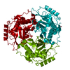
| Entry | Database: PDB / ID: 3nv7 | ||||||
|---|---|---|---|---|---|---|---|
| Title | Crystal structure of H.pylori phosphopantetheine adenylyltransferase mutant I4V/N76Y | ||||||
 Components Components | Phosphopantetheine adenylyltransferase | ||||||
 Keywords Keywords | TRANSFERASE / Helicobacter pylori 26695 strain / mutant I4V/N76Y / phosphopantetheine adenylyltransferase | ||||||
| Function / homology |  Function and homology information Function and homology informationpantetheine-phosphate adenylyltransferase / pantetheine-phosphate adenylyltransferase activity / coenzyme A biosynthetic process / ATP binding / cytoplasm Similarity search - Function | ||||||
| Biological species |   Helicobacter pylori (bacteria) Helicobacter pylori (bacteria) | ||||||
| Method |  X-RAY DIFFRACTION / SYNCHROTRON / MOLECULAR REPLACEMENT / Resolution: 1.75 Å X-RAY DIFFRACTION / SYNCHROTRON / MOLECULAR REPLACEMENT / Resolution: 1.75 Å | ||||||
 Authors Authors | Chen, C.H. / Cheng, C.S. / Yin, H.S. | ||||||
 Citation Citation | Journal: To be Published Title: Crystal structure of H.pylori phosphopantetheine adenylyltransferase mutant I4V/N76Y Authors: Chen, C.H. / Cheng, C.S. / Yin, H.S. | ||||||
| History |
|
- Structure visualization
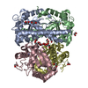
| Structure viewer | Molecule: MolmilJmol/JSmol |
|---|
- Downloads & links
-Download
| PDBx/mmCIF format | 3nv7.cif.gz | 81.8 KB | Display | PDBx/mmCIF format |
|---|---|---|---|---|
| PDB format | pdb3nv7.ent.gz | 61 KB | Display | PDB format |
| PDBx/mmJSON format | 3nv7.json.gz | Tree view | PDBx/mmJSON format | |
| Others |  Other downloads Other downloads |
-Validation report
| Arichive directory | https://data.pdbj.org/pub/pdb/validation_reports/nv/3nv7ftp://data.pdbj.org/pub/pdb/validation_reports/nv/3nv7 | HTTPS FTP |
|---|
-Related structure data
| Related structure data |  1h1tS S: Starting model for refinement |
|---|---|
| Similar structure data |


-Links
 PDBj
PDBj

- Assembly
| Deposited unit | 
| ||||||||
|---|---|---|---|---|---|---|---|---|---|
| 1 | 
| ||||||||
| Unit cell |
| ||||||||
| Components on special symmetry positions |
|
-Components
| #1: Protein | Mass: 17726.623 Da / Num. of mol.: 1 / Mutation: I4V, N76Y Source method: isolated from a genetically manipulated source Source: (gene. exp.) Helicobacter pylori (bacteria) / Strain: 26695 / Gene: coaD, kdtB, HP_1475 / Production host: References: UniProt: O26010, pantetheine-phosphate adenylyltransferase | ||||
|---|---|---|---|---|---|
| #2: Chemical | ChemComp-SO4 /   Mass: 96.063 Da / Num. of mol.: 4 / Source method: obtained synthetically / Formula: SO4 Mass: 96.063 Da / Num. of mol.: 4 / Source method: obtained synthetically / Formula: SO4#3: Chemical | ChemComp-ACY / |   Mass: 60.052 Da / Num. of mol.: 1 / Source method: obtained synthetically / Formula: C2H4O2 Mass: 60.052 Da / Num. of mol.: 1 / Source method: obtained synthetically / Formula: C2H4O2#4: Water | ChemComp-HOH / |  Mass: 18.015 Da / Num. of mol.: 136 / Source method: isolated from a natural source / Formula: H2O Mass: 18.015 Da / Num. of mol.: 136 / Source method: isolated from a natural source / Formula: H2O |
-Experimental details
-Experiment
| Experiment | Method: X-RAY DIFFRACTION / Number of used crystals: 1 |
|---|
- Sample preparation
| Crystal | Density Matthews: 2.54 Å3/Da / Density % sol: 51.54 % |
|---|---|
| Crystal grow | Temperature: 293 K / Method: evaporation / pH: 5.5 Details: 10% PEG 4000, 0.1M sodium acetate, 0.2M Li2SO4, pH 5.5, EVAPORATION, temperature 293K |
-Data collection
| Diffraction | Mean temperature: 100 K |
|---|---|
| Diffraction source | Source: SYNCHROTRON / Site: NSRRC  / Beamline: BL13B1 / Wavelength: 1 Å / Beamline: BL13B1 / Wavelength: 1 Å |
| Detector | Type: ADSC QUANTUM 315 / Detector: CCD / Date: Aug 22, 2008 |
| Radiation | Protocol: SINGLE WAVELENGTH / Monochromatic (M) / Laue (L): M / Scattering type: x-ray |
| Radiation wavelength | Wavelength: 1 Å / Relative weight: 1 |
| Reflection | Resolution: 1.75→30 Å / Num. obs: 18626 |
| Reflection shell | Resolution: 1.75→1.81 Å / % possible all: 98.6 |
- Processing
| Software |
| ||||||||||||||||||||||||||||||||||||||||||||||||||||||||||||||||||||||||||||||||||||||||||
|---|---|---|---|---|---|---|---|---|---|---|---|---|---|---|---|---|---|---|---|---|---|---|---|---|---|---|---|---|---|---|---|---|---|---|---|---|---|---|---|---|---|---|---|---|---|---|---|---|---|---|---|---|---|---|---|---|---|---|---|---|---|---|---|---|---|---|---|---|---|---|---|---|---|---|---|---|---|---|---|---|---|---|---|---|---|---|---|---|---|---|---|
| Refinement | Method to determine structure: MOLECULAR REPLACEMENT Starting model: 1H1T Resolution: 1.75→23.98 Å / Cor.coef. Fo:Fc: 0.958 / Cor.coef. Fo:Fc free: 0.939 / Occupancy max: 1 / Occupancy min: 0.5 / SU B: 5.788 / SU ML: 0.084 / Cross valid method: THROUGHOUT / σ(F): 0 / ESU R Free: 0.121 / Stereochemistry target values: MAXIMUM LIKELIHOOD / Details: HYDROGENS HAVE BEEN ADDED IN THE RIDING POSITIONS
| ||||||||||||||||||||||||||||||||||||||||||||||||||||||||||||||||||||||||||||||||||||||||||
| Solvent computation | Ion probe radii: 0.8 Å / Shrinkage radii: 0.8 Å / VDW probe radii: 1.2 Å / Solvent model: MASK | ||||||||||||||||||||||||||||||||||||||||||||||||||||||||||||||||||||||||||||||||||||||||||
| Displacement parameters | Biso max: 122.82 Å2 / Biso mean: 31.2635 Å2 / Biso min: 16.25 Å2
| ||||||||||||||||||||||||||||||||||||||||||||||||||||||||||||||||||||||||||||||||||||||||||
| Refinement step | Cycle: LAST / Resolution: 1.75→23.98 Å
| ||||||||||||||||||||||||||||||||||||||||||||||||||||||||||||||||||||||||||||||||||||||||||
| Refine LS restraints |
| ||||||||||||||||||||||||||||||||||||||||||||||||||||||||||||||||||||||||||||||||||||||||||
| LS refinement shell | Resolution: 1.749→1.794 Å / Total num. of bins used: 20
|
