 Movie
Movie Controller

[English] 日本語
 Yorodumi
- PDB-3nr0: Optimization of the in silico designed Kemp eliminase KE70 by com... -
+ Open data

- Basic information
| Entry | Database: PDB / ID: 3nr0 | ||||||
|---|---|---|---|---|---|---|---|
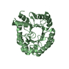
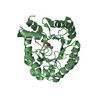
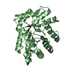
| Title | Optimization of the in silico designed Kemp eliminase KE70 by computational design and directed evolution R6 6/10A | ||||||
 Components Components | deoxyribose phosphate aldolase | ||||||
 Keywords Keywords | LYASE / TIM / Structural Genomics / Israel Structural Proteomics Center / ISPC | ||||||
| Function / homology | Aldolase class I / TIM Barrel / Alpha-Beta Barrel / Alpha Beta Function and homology information Function and homology information | ||||||
| Biological species |  | ||||||
| Method |  X-RAY DIFFRACTION / MOLECULAR REPLACEMENT / Resolution: 2.19 Å X-RAY DIFFRACTION / MOLECULAR REPLACEMENT / Resolution: 2.19 Å | ||||||
 Authors Authors | Khersonsky, O. / Rothlisberge, D. / Wollacott, A.M. / Dym, O. / Baker, D. / Tawfik, D.S. / Israel Structural Proteomics Center (ISPC) | ||||||
 Citation Citation | Journal: J.Mol.Biol. / Year: 2011 Title: Optimization of the in-silico-designed kemp eliminase KE70 by computational design and directed evolution Authors: Khersonsky, O. / Rothlisberger, D. / Wollacott, A.M. / Murphy, P. / Dym, O. / Albeck, S. / Kiss, G. / Houk, K.N. / Baker, D. / Tawfik, D.S. | ||||||
| History |
|
- Structure visualization
| Structure viewer | Molecule: MolmilJmol/JSmol |
|---|
- Downloads & links
-Download
| PDBx/mmCIF format | 3nr0.cif.gz | 102.1 KB | Display | PDBx/mmCIF format |
|---|---|---|---|---|
| PDB format | pdb3nr0.ent.gz | 78 KB | Display | PDB format |
| PDBx/mmJSON format | 3nr0.json.gz | Tree view | PDBx/mmJSON format | |
| Others |  Other downloads Other downloads |
-Validation report
| Arichive directory | https://data.pdbj.org/pub/pdb/validation_reports/nr/3nr0ftp://data.pdbj.org/pub/pdb/validation_reports/nr/3nr0 | HTTPS FTP |
|---|
-Related structure data
| Related structure data |  3npuSC  3npvC  3npwC  3npxC  3nq2C  3nq8C  3nqvC  3q2dC S: Starting model for refinement C: citing same article ( |
|---|---|
| Similar structure data |


-Links
 PDBj
PDBj- Assembly
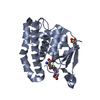
| Deposited unit | 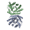
| ||||||||
|---|---|---|---|---|---|---|---|---|---|
| 1 | 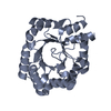
| ||||||||
| 2 | 
| ||||||||
| Unit cell |
|
-Components
| #1: Protein | Mass: 28803.760 Da / Num. of mol.: 2 Source method: isolated from a genetically manipulated source Source: (gene. exp.) #2: Water | ChemComp-HOH / |  Mass: 18.015 Da / Num. of mol.: 66 / Source method: isolated from a natural source / Formula: H2O Mass: 18.015 Da / Num. of mol.: 66 / Source method: isolated from a natural source / Formula: H2OSequence details | A SEQUENCE DATABASE REFERENCE FOR THIS PROTEIN DOES NOT CURRENTLY EXIST. THE WILD TYPE HAS BEEN ...A SEQUENCE DATABASE REFERENCE FOR THIS PROTEIN DOES NOT CURRENTLY EXIST. THE WILD TYPE HAS BEEN DEPOSITED TO PDB, 3NPU AND 3NPV. THIS SEQUENCE IS ALA 2, SER 19A, ALA 239 INSERTION AND K28N, T42N, Y47F, W71C, G100S, S137A, H165N, A203V MUTANT. ALSO ASP RESIDUE BETWEEN THR 3 AND LEU 4 HAS BEEN DELETION. | |
|---|
-Experimental details
-Experiment
| Experiment | Method: X-RAY DIFFRACTION / Number of used crystals: 1 |
|---|
- Sample preparation
| Crystal | Density Matthews: 2.44 Å3/Da / Density % sol: 49.6 % |
|---|---|
| Crystal grow | Temperature: 292 K / Method: vapor diffusion, sitting drop / pH: 5 Details: 0.2M MgCl2, 0.1M NaAc, 20% PEG 6000, pH 5, VAPOR DIFFUSION, SITTING DROP, temperature 292K |
-Data collection
| Diffraction source | Source: ROTATING ANODE / Type: RIGAKU RU300 / Wavelength: 1.5418 Å |
|---|---|
| Detector | Type: RIGAKU RAXIS IV++ / Detector: IMAGE PLATE / Date: Jul 9, 2008 / Details: Mirrors |
| Radiation | Protocol: SINGLE WAVELENGTH / Monochromatic (M) / Laue (L): M / Scattering type: x-ray |
| Radiation wavelength | Wavelength: 1.5418 Å / Relative weight: 1 |
| Reflection | Resolution: 2.19→50 Å / Num. all: 29029 / Num. obs: 27694 / % possible obs: 95.4 % / Redundancy: 4 % / Biso Wilson estimate: 31.4 Å2 / Rmerge(I) obs: 0.098 / Rsym value: 0.073 / Net I/σ(I): 15.5 |
| Reflection shell | Resolution: 2.2→2.24 Å / Redundancy: 4 % / Rmerge(I) obs: 0.49 / Mean I/σ(I) obs: 3.08 / Num. unique all: 1383 / Rsym value: 0.43 / % possible all: 95.7 |
- Processing
| Software |
| ||||||||||||||||||||||||||||||||||||||||||||||||||||||||||||||||||||||||||||||||||||||||||||||||||||||||||||||||||||||||||||||||||||||||||||||||||||||||||||||||||||||||||
|---|---|---|---|---|---|---|---|---|---|---|---|---|---|---|---|---|---|---|---|---|---|---|---|---|---|---|---|---|---|---|---|---|---|---|---|---|---|---|---|---|---|---|---|---|---|---|---|---|---|---|---|---|---|---|---|---|---|---|---|---|---|---|---|---|---|---|---|---|---|---|---|---|---|---|---|---|---|---|---|---|---|---|---|---|---|---|---|---|---|---|---|---|---|---|---|---|---|---|---|---|---|---|---|---|---|---|---|---|---|---|---|---|---|---|---|---|---|---|---|---|---|---|---|---|---|---|---|---|---|---|---|---|---|---|---|---|---|---|---|---|---|---|---|---|---|---|---|---|---|---|---|---|---|---|---|---|---|---|---|---|---|---|---|---|---|---|---|---|---|---|---|
| Refinement | Method to determine structure: MOLECULAR REPLACEMENT Starting model: 3NPU Resolution: 2.19→50 Å / Cor.coef. Fo:Fc: 0.936 / Cor.coef. Fo:Fc free: 0.905 / SU B: 6.321 / SU ML: 0.161 / Cross valid method: THROUGHOUT / ESU R Free: 0.229 / Stereochemistry target values: MAXIMUM LIKELIHOOD / Details: HYDROGENS HAVE BEEN ADDED IN THE RIDING POSITIONS
| ||||||||||||||||||||||||||||||||||||||||||||||||||||||||||||||||||||||||||||||||||||||||||||||||||||||||||||||||||||||||||||||||||||||||||||||||||||||||||||||||||||||||||
| Solvent computation | Ion probe radii: 0.8 Å / Shrinkage radii: 0.8 Å / VDW probe radii: 1.2 Å / Solvent model: MASK | ||||||||||||||||||||||||||||||||||||||||||||||||||||||||||||||||||||||||||||||||||||||||||||||||||||||||||||||||||||||||||||||||||||||||||||||||||||||||||||||||||||||||||
| Displacement parameters | Biso mean: 31.412 Å2
| ||||||||||||||||||||||||||||||||||||||||||||||||||||||||||||||||||||||||||||||||||||||||||||||||||||||||||||||||||||||||||||||||||||||||||||||||||||||||||||||||||||||||||
| Refinement step | Cycle: LAST / Resolution: 2.19→50 Å
| ||||||||||||||||||||||||||||||||||||||||||||||||||||||||||||||||||||||||||||||||||||||||||||||||||||||||||||||||||||||||||||||||||||||||||||||||||||||||||||||||||||||||||
| Refine LS restraints |
| ||||||||||||||||||||||||||||||||||||||||||||||||||||||||||||||||||||||||||||||||||||||||||||||||||||||||||||||||||||||||||||||||||||||||||||||||||||||||||||||||||||||||||
| LS refinement shell | Resolution: 2.19→2.247 Å / Total num. of bins used: 20
|
