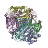
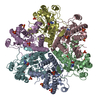
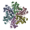
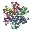
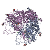
- PDB-3no4: Crystal structure of a creatinine amidohydrolase (Npun_F1913) fro... -
+
Open data
ID or keywords:
Loading...
-
Basic information
Entry
Database: PDB / ID: 3no4
Title
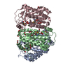
Crystal structure of a creatinine amidohydrolase (Npun_F1913) from Nostoc punctiforme PCC 73102 at 2.00 A resolution
Components
creatinine amidohydrolase
Keywords
HYDROLASE / STRUCTURAL GENOMICS / JOINT CENTER FOR STRUCTURAL GENOMICS / JCSG / PROTEIN STRUCTURE INITIATIVE / PSI-2
Function / homology
Function and homology information
hydrolase activity, acting on carbon-nitrogen (but not peptide) bonds, in linear amides / riboflavin biosynthetic process / metal ion binding Similarity search - Function
Component-ID: 1 / Ens-ID: 1 / Beg auth comp-ID: MSE / Beg label comp-ID: MSE / End auth comp-ID: ASP / End label comp-ID: ASP / Refine code: 3 / Auth seq-ID: 1 - 248 / Label seq-ID: 20 - 267
Dom-ID
Auth asym-ID
Label asym-ID
1
A
A
2
B
B
3
C
C
Details
CRYSTAL PACKING ANALYSIS SUGGESTS THE ASSIGNMENT OF A HEXAMER AS THE SIGNIFICANT OLIGOMERIZATION STATE. HOWEVER, SIZE EXCLUSION CHROMATOGRAPHY ALONG WITH STATIC LIGHT SCATTERING EXPERIMENT SUGGESTS THE ASSIGNMENT OF A PENTAMER AS THE SIGNIFICANT OLIGOMERIZATION STATE.
-
Components
-
Protein , 1 types, 3 molecules ABC
#1: Protein
creatinineamidohydrolase / Creatininase
Mass: 29241.070 Da / Num. of mol.: 3 Source method: isolated from a genetically manipulated source Source: (gene. exp.) Nostoc punctiforme PCC 73102 (bacteria) Gene: NPUN_22DEC03_CONTIG1_REVISED_GENENPF1913, Npun_F1913 / Plasmid: SpeedET / Production host: Escherichia Coli (E. coli) / Strain (production host): HK100 / References: UniProt: B2J3U5
Type: MARMOSAIC 325 mm CCD / Detector: CCD / Date: Dec 3, 2009 / Details: FLAT MIRROR (VERTICAL FOCUSING)
Radiation
Monochromator: SINGLE CRYSTAL SI(111) BENT MONOCHROMATOR (HORIZONTAL FOCUSING) Protocol: MAD / Monochromatic (M) / Laue (L): M / Scattering type: x-ray
Radiation wavelength
ID
Wavelength (Å)
Relative weight
1
0.91837
1
2
0.97939
1
3
0.97901
1
Reflection
Resolution: 2→29.695 Å / Num. obs: 58475 / % possible obs: 99.9 % / Redundancy: 7.3 % / Biso Wilson estimate: 20.76 Å2 / Rsym value: 0.164 / Net I/σ(I): 9.9
Reflection shell
Resolution (Å)
Rmerge(I) obs
Mean I/σ(I) obs
Num. measured obs
Num. unique obs
% possible all
2-2.05
0.943
2.4
31503
4244
99.9
2.05-2.11
0.788
2.9
30771
4135
99.9
2.11-2.17
0.638
3.5
29991
4033
99.9
2.17-2.24
0.516
4.2
29054
3909
99.9
2.24-2.31
0.469
4.6
28142
3791
100
2.31-2.39
0.402
5.3
27337
3685
100
2.39-2.48
0.338
6.1
26546
3685
100
2.48-2.58
0.304
6.6
25421
3574
100
2.58-2.7
0.236
8.3
24463
3432
100
2.7-2.83
0.215
8.9
23356
3303
100
2.83-2.98
0.166
10.9
22400
3157
100
2.98-3.16
0.132
13.1
21158
3043
100
3.16-3.38
0.11
15.2
19766
2688
100
3.38-3.65
0.092
18.1
18425
2516
100
3.65-4
0.077
21.7
17034
2343
100
4-4.47
0.063
24.8
15489
2147
100
4.47-5.16
0.06
24.6
13543
1894
100
5.16-6.32
0.067
20.7
11606
1650
100
6.32-8.94
0.056
23.8
8895
1300
100
8.94-29.7
0.048
24.7
4719
765
97.4
-
Phasing
Phasing
Method: MAD
-
Processing
Software
Name
Version
Classification
NB
REFMAC
5.5.0110
refinement
PHENIX
refinement
SOLVE
phasing
MolProbity
3beta29
modelbuilding
SCALA
3.3.15
datascaling
PDB_EXTRACT
3.006
dataextraction
MOSFLM
datareduction
Refinement
Method to determine structure: MAD / Resolution: 2→29.695 Å / Cor.coef. Fo:Fc: 0.965 / Cor.coef. Fo:Fc free: 0.948 / Occupancy max: 1 / Occupancy min: 0.23 / SU B: 6.437 / SU ML: 0.091 / Cross valid method: THROUGHOUT / σ(F): 0 / ESU R Free: 0.136 Stereochemistry target values: MAXIMUM LIKELIHOOD WITH PHASES Details: 1. HYDROGENS HAVE BEEN ADDED IN THE RIDING POSITIONS. 2. ATOM RECORD CONTAINS SUM OF TLS AND RESIDUAL B FACTORS. 3. ANISOU RECORD CONTAINS SUM OF TLS AND RESIDUAL U FACTORS. 4. WATERS WERE ...Details: 1. HYDROGENS HAVE BEEN ADDED IN THE RIDING POSITIONS. 2. ATOM RECORD CONTAINS SUM OF TLS AND RESIDUAL B FACTORS. 3. ANISOU RECORD CONTAINS SUM OF TLS AND RESIDUAL U FACTORS. 4. WATERS WERE EXCLUDED FROM AUTOMATIC TLS ASSIGNMENT. 5. A MET-INHIBITION PROTOCOL WAS USED FOR SELENOMETHIONINE INCORPORATION DURING PROTEIN EXPRESSION. THE OCCUPANCY OF THE SE ATOMS IN THE MSE RESIDUES WAS REDUCED TO 0.75 FOR THE REDUCED SCATTERING POWER DUE TO PARTIAL S-MET INCORPORATION. 6. X-RAY FLUORESCENCE EXCITATION AND WAVELENGTH SCANS AND ANOMALOUS DIFFERENCE FOURIERS SUPPORT THE MODELING OF NI IONS. HOWEVER, TRACE AMOUNTS OF ZN MAY BE PRESENT AT THESE SITES. 7. ETHYLENE GLYCOL (EDO) MOLECULES FROM THE CRYOPROTECTION SOLUTION ARE MODELED. 8. AN UNKNOWN LIGAND MOLECULE (UNL) IS MODELED NEAR THE NI ION IN THE ACTIVE SITE IN EACH CHAIN.
Rfactor
Num. reflection
% reflection
Selection details
Rfree
0.192
2955
5.1 %
RANDOM
Rwork
0.151
-
-
-
obs
0.153
58399
100 %
-
Solvent computation
Ion probe radii: 0.8 Å / Shrinkage radii: 0.8 Å / VDW probe radii: 1.4 Å / Solvent model: BABINET MODEL WITH MASK
Displacement parameters
Biso mean: 28.72 Å2
Baniso -1
Baniso -2
Baniso -3
1-
1.15 Å2
0 Å2
0 Å2
2-
-
1.15 Å2
0 Å2
3-
-
-
-2.3 Å2
Refinement step
Cycle: LAST / Resolution: 2→29.695 Å
Protein
Nucleic acid
Ligand
Solvent
Total
Num. atoms
5863
0
76
581
6520
Refine LS restraints
Refine-ID
Type
Dev ideal
Dev ideal target
Number
X-RAY DIFFRACTION
r_bond_refined_d
0.017
0.022
6269
X-RAY DIFFRACTION
r_bond_other_d
0.003
0.02
4127
X-RAY DIFFRACTION
r_angle_refined_deg
1.536
1.946
8539
X-RAY DIFFRACTION
r_angle_other_deg
1.306
3
10136
X-RAY DIFFRACTION
r_dihedral_angle_1_deg
6.297
5
829
X-RAY DIFFRACTION
r_dihedral_angle_2_deg
36.522
25.017
289
X-RAY DIFFRACTION
r_dihedral_angle_3_deg
13.051
15
973
X-RAY DIFFRACTION
r_dihedral_angle_4_deg
17.489
15
18
X-RAY DIFFRACTION
r_chiral_restr
0.092
0.2
932
X-RAY DIFFRACTION
r_gen_planes_refined
0.007
0.021
7163
X-RAY DIFFRACTION
r_gen_planes_other
0.002
0.02
1241
X-RAY DIFFRACTION
r_nbd_refined
X-RAY DIFFRACTION
r_nbd_other
X-RAY DIFFRACTION
r_nbtor_refined
X-RAY DIFFRACTION
r_nbtor_other
X-RAY DIFFRACTION
r_xyhbond_nbd_refined
X-RAY DIFFRACTION
r_xyhbond_nbd_other
X-RAY DIFFRACTION
r_metal_ion_refined
X-RAY DIFFRACTION
r_metal_ion_other
X-RAY DIFFRACTION
r_symmetry_vdw_refined
X-RAY DIFFRACTION
r_symmetry_vdw_other
X-RAY DIFFRACTION
r_symmetry_hbond_refined
X-RAY DIFFRACTION
r_symmetry_hbond_other
X-RAY DIFFRACTION
r_symmetry_metal_ion_refined
X-RAY DIFFRACTION
r_symmetry_metal_ion_other
X-RAY DIFFRACTION
r_mcbond_it
0.685
1.5
3907
X-RAY DIFFRACTION
r_mcbond_other
0.204
1.5
1608
X-RAY DIFFRACTION
r_mcangle_it
1.188
2
6283
X-RAY DIFFRACTION
r_scbond_it
2.078
3
2362
X-RAY DIFFRACTION
r_scangle_it
3.217
4.5
2224
X-RAY DIFFRACTION
r_rigid_bond_restr
X-RAY DIFFRACTION
r_sphericity_free
X-RAY DIFFRACTION
r_sphericity_bonded
Refine LS restraints NCS
Ens-ID: 1 / Refine-ID: X-RAY DIFFRACTION
Dom-ID
Auth asym-ID
Number
Type
Rms dev position (Å)
Weight position
1
A
1438
tightpositional
0.12
0.05
2
B
1438
tightpositional
0.12
0.05
3
C
1438
tightpositional
0.11
0.05
1
A
1597
loosepositional
0.24
5
2
B
1597
loosepositional
0.31
5
3
C
1597
loosepositional
0.27
5
1
A
1438
tightthermal
1.25
0.5
2
B
1438
tightthermal
1.19
0.5
3
C
1438
tightthermal
1.36
0.5
1
A
1597
loosethermal
1.61
10
2
B
1597
loosethermal
1.5
10
3
C
1597
loosethermal
1.51
10
LS refinement shell
Resolution: 2→2.05 Å / Total num. of bins used: 20
Rfactor
Num. reflection
% reflection
Rfree
0.277
209
-
Rwork
0.221
4011
-
obs
-
-
99.81 %
Refinement TLS params.
Method: refined / Refine-ID: X-RAY DIFFRACTION
ID
L11 (°2)
L12 (°2)
L13 (°2)
L22 (°2)
L23 (°2)
L33 (°2)
S11 (Å °)
S12 (Å °)
S13 (Å °)
S21 (Å °)
S22 (Å °)
S23 (Å °)
S31 (Å °)
S32 (Å °)
S33 (Å °)
T11 (Å2)
T12 (Å2)
T13 (Å2)
T22 (Å2)
T23 (Å2)
T33 (Å2)
Origin x (Å)
Origin y (Å)
Origin z (Å)
1
0.5238
-0.1644
0.0508
0.8988
-0.0213
1.1316
-0.0157
-0.0154
0.1516
0.0365
0.0501
-0.0979
-0.2614
0.0494
-0.0343
0.0718
-0.0115
0.0255
0.0122
-0.01
0.1327
97.6199
30.1262
3.9415
2
0.4867
-0.1191
-0.0071
0.6336
0.1554
1.1483
-0.0002
-0.0094
0.0669
0.0025
-0.0012
0.1297
-0.1648
-0.2418
0.0014
0.0321
0.0341
0.0228
0.0594
0.0006
0.121
75.3035
23.1327
7.2642
3
0.3813
-0.0057
-0.2275
0.3629
-0.1351
1.3095
0.0155
0.0681
-0.0314
0.0254
0.0159
0.1236
0.057
-0.3026
-0.0314
0.0272
-0.0367
0.009
0.0987
0.0021
0.1231
67.5714
-4.7498
2.7177
Refinement TLS group
ID
Refine-ID
Refine TLS-ID
Auth asym-ID
Auth seq-ID
1
X-RAY DIFFRACTION
1
A
-15 - 248
2
X-RAY DIFFRACTION
1
A
300
3
X-RAY DIFFRACTION
2
B
-7 - 248
4
X-RAY DIFFRACTION
2
B
300
5
X-RAY DIFFRACTION
3
C
-1 - 248
6
X-RAY DIFFRACTION
3
C
300
+
About Yorodumi
-
News
-
Feb 9, 2022. New format data for meta-information of EMDB entries
New format data for meta-information of EMDB entries
Version 3 of the EMDB header file is now the official format.
The previous official version 1.9 will be removed from the archive.
In the structure databanks used in Yorodumi, some data are registered as the other names, "COVID-19 virus" and "2019-nCoV". Here are the details of the virus and the list of structure data.
Jan 31, 2019. EMDB accession codes are about to change! (news from PDBe EMDB page)
EMDB accession codes are about to change! (news from PDBe EMDB page)
The allocation of 4 digits for EMDB accession codes will soon come to an end. Whilst these codes will remain in use, new EMDB accession codes will include an additional digit and will expand incrementally as the available range of codes is exhausted. The current 4-digit format prefixed with “EMD-” (i.e. EMD-XXXX) will advance to a 5-digit format (i.e. EMD-XXXXX), and so on. It is currently estimated that the 4-digit codes will be depleted around Spring 2019, at which point the 5-digit format will come into force.
The EM Navigator/Yorodumi systems omit the EMD- prefix.
Related info.:Q: What is EMD? / ID/Accession-code notation in Yorodumi/EM Navigator
Yorodumi is a browser for structure data from EMDB, PDB, SASBDB, etc.
This page is also the successor to EM Navigator detail page, and also detail information page/front-end page for Omokage search.
The word "yorodu" (or yorozu) is an old Japanese word meaning "ten thousand". "mi" (miru) is to see.
Related info.:EMDB / PDB / SASBDB / Comparison of 3 databanks / Yorodumi Search / Aug 31, 2016. New EM Navigator & Yorodumi / Yorodumi Papers / Jmol/JSmol / Function and homology information / Changes in new EM Navigator and Yorodumi
 Movie
Movie Controller

 Yorodumi
 Open data
 Basic information
 Components
 Keywords
 Function and homology information
 Nostoc punctiforme PCC 73102 (bacteria)
 X-RAY DIFFRACTION /
  Authors
 Citation
 Structure visualization
 Downloads & links
 Other downloads
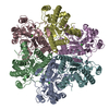

 PDBj
PDBj

 Assembly
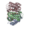


 Mass: 58.693 Da / Num. of mol.: 3 / Source method: obtained synthetically / Formula: Ni
 Mass: 35.453 Da / Num. of mol.: 1 / Source method: obtained synthetically / Formula: Cl
 Mass: 62.068 Da / Num. of mol.: 16 / Source method: obtained synthetically / Formula: C2H6O2
 Sample preparation
 / Beamline: BL11-1 / Wavelength: 0.91837,0.97939,0.97901
 Processing