 Movie
Movie Controller


+ Open data

- Basic information
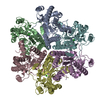
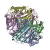
| Entry | Database: PDB / ID: 3a6f | ||||||
|---|---|---|---|---|---|---|---|
| Title | W174F mutant creatininase, Type II | ||||||
 Components Components | Creatinine amidohydrolase | ||||||
 Keywords Keywords | HYDROLASE / creatinine amidohydrolase / urease-related amidohydrolase superfamily | ||||||
| Function / homology |  Function and homology information Function and homology informationcreatininase / creatininase activity / creatinine catabolic process / creatine biosynthetic process / hydrolase activity, acting on carbon-nitrogen (but not peptide) bonds, in linear amides / riboflavin biosynthetic process / manganese ion binding / zinc ion binding Similarity search - Function | ||||||
| Biological species |  Pseudomonas putida (bacteria) Pseudomonas putida (bacteria) | ||||||
| Method |  X-RAY DIFFRACTION / SYNCHROTRON / MOLECULAR REPLACEMENT / Resolution: 1.78 Å X-RAY DIFFRACTION / SYNCHROTRON / MOLECULAR REPLACEMENT / Resolution: 1.78 Å | ||||||
 Authors Authors | Nakajima, Y. / Yamashita, K. / Ito, K. / Yoshimoto, T. | ||||||
 Citation Citation | Journal: J.Mol.Biol. / Year: 2010 Title: Substitution of Glu122 by glutamine revealed the function of the second water molecule as a proton donor in the binuclear metal enzyme creatininase Authors: Yamashita, K. / Nakajima, Y. / Matsushita, H. / Nishiya, Y. / Yamazawa, R. / Wu, Y.F. / Matsubara, F. / Oyama, H. / Ito, K. / Yoshimoto, T. | ||||||
| History |
|
- Structure visualization
| Structure viewer | Molecule: MolmilJmol/JSmol |
|---|
- Downloads & links
-Download
| PDBx/mmCIF format | 3a6f.cif.gz | 315.7 KB | Display | PDBx/mmCIF format |
|---|---|---|---|---|
| PDB format | pdb3a6f.ent.gz | 256.5 KB | Display | PDB format |
| PDBx/mmJSON format | 3a6f.json.gz | Tree view | PDBx/mmJSON format | |
| Others |  Other downloads Other downloads |
-Validation report
| Arichive directory | https://data.pdbj.org/pub/pdb/validation_reports/a6/3a6fftp://data.pdbj.org/pub/pdb/validation_reports/a6/3a6f | HTTPS FTP |
|---|
-Related structure data
| Related structure data | 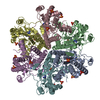 3a6dC 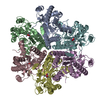 3a6eC  3a6gC  3a6hC 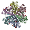 3a6jC 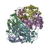 3a6kC  3a6lC  1j2tS S: Starting model for refinement C: citing same article ( |
|---|---|
| Similar structure data |


-Links
 PDBj
PDBj


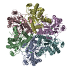
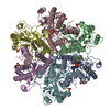
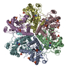
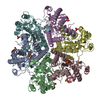
- Assembly
| Deposited unit | 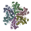
| ||||||||
|---|---|---|---|---|---|---|---|---|---|
| 1 |
| ||||||||
| Unit cell |
|
-Components
| #1: Protein | Mass: 28559.754 Da / Num. of mol.: 6 / Mutation: W174F Source method: isolated from a genetically manipulated source Source: (gene. exp.) Pseudomonas putida (bacteria) / Plasmid: pUC19 / Production host: #2: Chemical | ChemComp-MN /   Mass: 54.938 Da / Num. of mol.: 6 / Source method: obtained synthetically / Formula: Mn Mass: 54.938 Da / Num. of mol.: 6 / Source method: obtained synthetically / Formula: Mn#3: Chemical | ChemComp-ZN /   Mass: 65.409 Da / Num. of mol.: 6 / Source method: obtained synthetically / Formula: Zn Mass: 65.409 Da / Num. of mol.: 6 / Source method: obtained synthetically / Formula: Zn#4: Chemical | ChemComp-CAC /   Mass: 136.989 Da / Num. of mol.: 6 / Source method: obtained synthetically / Formula: C2H6AsO2 Mass: 136.989 Da / Num. of mol.: 6 / Source method: obtained synthetically / Formula: C2H6AsO2#5: Water | ChemComp-HOH / |  Mass: 18.015 Da / Num. of mol.: 764 / Source method: isolated from a natural source / Formula: H2O Mass: 18.015 Da / Num. of mol.: 764 / Source method: isolated from a natural source / Formula: H2O |
|---|
-Experimental details
-Experiment
| Experiment | Method: X-RAY DIFFRACTION / Number of used crystals: 1 |
|---|
- Sample preparation
| Crystal | Density Matthews: 3.73 Å3/Da / Density % sol: 66.99 % |
|---|---|
| Crystal grow | Temperature: 293 K / Method: vapor diffusion, hanging drop / pH: 6 Details: 45% PEG200, 20mM Magnesium chloride, 100mM Sodium cacodylate buffer, pH 6.0, VAPOR DIFFUSION, HANGING DROP, temperature 293K |
-Data collection
| Diffraction | Mean temperature: 100 K |
|---|---|
| Diffraction source | Source: SYNCHROTRON / Site: Photon Factory  / Beamline: BL-5A / Wavelength: 1 Å / Beamline: BL-5A / Wavelength: 1 Å |
| Detector | Type: ADSC QUANTUM 315r / Detector: CCD / Date: May 10, 2006 / Details: Rhodium coated silicon single crystal |
| Radiation | Monochromator: Numerical link type Si(111) double crystal monochromator Protocol: SINGLE WAVELENGTH / Monochromatic (M) / Laue (L): M / Scattering type: x-ray |
| Radiation wavelength | Wavelength: 1 Å / Relative weight: 1 |
| Reflection | Resolution: 1.78→50 Å / Num. all: 242993 / Num. obs: 242993 / % possible obs: 99.9 % / Observed criterion σ(F): 0 / Observed criterion σ(I): 0 / Redundancy: 11.1 % / Biso Wilson estimate: 23.5 Å2 / Rmerge(I) obs: 0.079 / Net I/σ(I): 41.8 |
| Reflection shell | Resolution: 1.78→1.84 Å / Redundancy: 10 % / Rmerge(I) obs: 0.242 / Mean I/σ(I) obs: 5.6 / Num. unique all: 24075 / % possible all: 99.8 |
- Processing
| Software |
| |||||||||||||||||||||||||
|---|---|---|---|---|---|---|---|---|---|---|---|---|---|---|---|---|---|---|---|---|---|---|---|---|---|---|
| Refinement | Method to determine structure: MOLECULAR REPLACEMENT Starting model: 1J2T Resolution: 1.78→20 Å / Isotropic thermal model: isotropic / Cross valid method: THROUGHOUT / σ(F): 0 / σ(I): 0 / Stereochemistry target values: Engh & Huber Details: The diffraction data was detwined with twin fraction of 0.463
| |||||||||||||||||||||||||
| Displacement parameters | Biso mean: 25 Å2 | |||||||||||||||||||||||||
| Refine analyze |
| |||||||||||||||||||||||||
| Refinement step | Cycle: LAST / Resolution: 1.78→20 Å
| |||||||||||||||||||||||||
| Refine LS restraints |
| |||||||||||||||||||||||||
| LS refinement shell | Resolution: 1.78→1.84 Å
|
