 Movie
Movie Controller

[English] 日本語
 Yorodumi
- PDB-3nnr: Crystal structure of a TetR-family transcriptional regulator (Maq... -
+ Open data

- Basic information
| Entry | Database: PDB / ID: 3nnr | ||||||
|---|---|---|---|---|---|---|---|
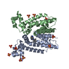
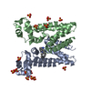
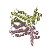
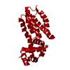
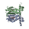
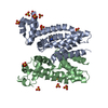
| Title | Crystal structure of a TetR-family transcriptional regulator (Maqu_3571) from MARINOBACTER AQUAEOLEI VT8 at 2.49 A resolution | ||||||
 Components Components | Transcriptional regulator, TetR family | ||||||
 Keywords Keywords | TRANSCRIPTION / TETR-FAMILY TRANSCRIPTIONAL REGULATOR / Structural Genomics / Joint Center for Structural Genomics / JCSG / Protein Structure Initiative / PSI-2 | ||||||
| Function / homology |  Function and homology information Function and homology information | ||||||
| Biological species |  Marinobacter aquaeolei (bacteria) Marinobacter aquaeolei (bacteria) | ||||||
| Method |  X-RAY DIFFRACTION / SYNCHROTRON / MAD / Resolution: 2.49 Å X-RAY DIFFRACTION / SYNCHROTRON / MAD / Resolution: 2.49 Å | ||||||
 Authors Authors | Joint Center for Structural Genomics (JCSG) | ||||||
 Citation Citation | Journal: To be published Title: Crystal structure of a TetR-family transcriptional regulator (Maqu_3571) from MARINOBACTER AQUAEOLEI VT8 at 2.49 A resolution Authors: Joint Center for Structural Genomics (JCSG) | ||||||
| History |
|
- Structure visualization
| Structure viewer | Molecule: MolmilJmol/JSmol |
|---|
- Downloads & links
-Download
| PDBx/mmCIF format | 3nnr.cif.gz | 102.3 KB | Display | PDBx/mmCIF format |
|---|---|---|---|---|
| PDB format | pdb3nnr.ent.gz | 78.7 KB | Display | PDB format |
| PDBx/mmJSON format | 3nnr.json.gz | Tree view | PDBx/mmJSON format | |
| Others |  Other downloads Other downloads |
-Validation report
| Arichive directory | https://data.pdbj.org/pub/pdb/validation_reports/nn/3nnrftp://data.pdbj.org/pub/pdb/validation_reports/nn/3nnr | HTTPS FTP |
|---|
-Related structure data
| Similar structure data | |
|---|---|
| Other databases |

-Links
 PDBj
PDBj

- Assembly
| Deposited unit | 
| ||||||||
|---|---|---|---|---|---|---|---|---|---|
| 1 | 
| ||||||||
| Unit cell |
| ||||||||
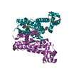
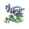
| Details | CRYSTAL PACKING ANALYSIS SUGGEST THE ASSIGNMENT OF A DIMER AS THE SIGNIFICANT OLIGOMERIZATION STATE IN SOLUTION. |
-Components
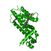
| #1: Protein | Mass: 26902.824 Da / Num. of mol.: 1 Source method: isolated from a genetically manipulated source Source: (gene. exp.) Marinobacter aquaeolei (bacteria) / Strain: ATCC 700491 / DSM 11845 / VT8 / Gene: Maqu_3571 / Plasmid: SpeedET / Production host: | ||||||
|---|---|---|---|---|---|---|---|
| #2: Chemical | ChemComp-UNL / Num. of mol.: 1 / Source method: obtained synthetically | ||||||
| #3: Chemical |   Mass: 96.063 Da / Num. of mol.: 2 / Source method: obtained synthetically / Formula: SO4 Mass: 96.063 Da / Num. of mol.: 2 / Source method: obtained synthetically / Formula: SO4#4: Water | ChemComp-HOH / |  Mass: 18.015 Da / Num. of mol.: 14 / Source method: isolated from a natural source / Formula: H2O Mass: 18.015 Da / Num. of mol.: 14 / Source method: isolated from a natural source / Formula: H2OHas protein modification | Y | Sequence details | THIS PROTEIN WAS EXPRESSED WITH A PURIFICATION TAG MGSDKIHHHHHHENLYFQG. THE TAG WAS REMOVED WITH ...THIS PROTEIN WAS EXPRESSED WITH A PURIFICATI | |
-Experimental details
-Experiment
| Experiment | Method: X-RAY DIFFRACTION / Number of used crystals: 1 |
|---|
- Sample preparation
| Crystal | Density Matthews: 2.77 Å3/Da / Density % sol: 55.57 % |
|---|---|
| Crystal grow | Temperature: 293 K / pH: 9.5 Details: 0.2000M lithium sulfate, 1.0500M potassium sodium tartrate, 0.1M CHES pH 9.5, NANODROP, VAPOR DIFFUSION, SITTING DROP, temperature 293K |
-Data collection
| Diffraction | Mean temperature: 100 K | ||||||||||||
|---|---|---|---|---|---|---|---|---|---|---|---|---|---|
| Diffraction source | Source: SYNCHROTRON / Site: SSRL  / Beamline: BL9-2 / Wavelength: 0.91837,0.97934,0.97922 / Beamline: BL9-2 / Wavelength: 0.91837,0.97934,0.97922 | ||||||||||||
| Detector | Type: MARMOSAIC 325 mm CCD / Detector: CCD / Date: May 14, 2010 / Details: FLAT COLLIMATING MIRROR, TOROID FOCUSING MIRROR | ||||||||||||
| Radiation | Monochromator: DOUBLE CRYSTAL MONOCHROMATOR / Protocol: MAD / Monochromatic (M) / Laue (L): M / Scattering type: x-ray | ||||||||||||
| Radiation wavelength |
| ||||||||||||
| Reflection | Resolution: 2.49→29.93 Å / Num. obs: 11338 / % possible obs: 100 % / Redundancy: 9.1 % / Biso Wilson estimate: 68.12 Å2 / Rsym value: 0.074 / Net I/σ(I): 16.9 | ||||||||||||
| Reflection shell | Highest resolution: 2.49 Å / Redundancy: 9.4 % / Mean I/σ(I) obs: 2.1 / Rsym value: 1.187 / % possible all: 100 |
-Phasing
| Phasing | Method: MAD |
|---|
- Processing
| Software |
| ||||||||||||||||||||||||||||||||||||||||||||||||||||||||||||||||||||||||||||||||||||||||||||||||||||||||||||||||||||||||||||||||||||||||||||||||||||||||||||||||||||||||||
|---|---|---|---|---|---|---|---|---|---|---|---|---|---|---|---|---|---|---|---|---|---|---|---|---|---|---|---|---|---|---|---|---|---|---|---|---|---|---|---|---|---|---|---|---|---|---|---|---|---|---|---|---|---|---|---|---|---|---|---|---|---|---|---|---|---|---|---|---|---|---|---|---|---|---|---|---|---|---|---|---|---|---|---|---|---|---|---|---|---|---|---|---|---|---|---|---|---|---|---|---|---|---|---|---|---|---|---|---|---|---|---|---|---|---|---|---|---|---|---|---|---|---|---|---|---|---|---|---|---|---|---|---|---|---|---|---|---|---|---|---|---|---|---|---|---|---|---|---|---|---|---|---|---|---|---|---|---|---|---|---|---|---|---|---|---|---|---|---|---|---|---|
| Refinement | Method to determine structure: MAD / Resolution: 2.49→29.93 Å / Cor.coef. Fo:Fc: 0.943 / Cor.coef. Fo:Fc free: 0.933 / Occupancy max: 1 / Occupancy min: 0.37 / SU B: 20.893 / SU ML: 0.227 / Cross valid method: THROUGHOUT / σ(F): 0 / ESU R Free: 0.265 Stereochemistry target values: MAXIMUM LIKELIHOOD WITH PHASES Details: 1.HYDROGENS HAVE BEEN ADDED IN THE RIDING POSITIONS. 2.A MET-INHIBITION PROTOCOL WAS USED FOR SELENOMETHIONINE INCORPORATION DURING PROTEIN EXPRESSION. THE OCCUPANCY OF THE SE ATOMS IN THE ...Details: 1.HYDROGENS HAVE BEEN ADDED IN THE RIDING POSITIONS. 2.A MET-INHIBITION PROTOCOL WAS USED FOR SELENOMETHIONINE INCORPORATION DURING PROTEIN EXPRESSION. THE OCCUPANCY OF THE SE ATOMS IN THE MSE RESIDUES WAS REDUCED TO 0.75 FOR THE REDUCED SCATTERING POWER DUE TO PARTIAL S-MET INCORPORATION. 3.ATOM RECORD CONTAINS SUM OF TLS AND RESIDUAL B FACTORS. ANISOU RECORD CONTAINS SUM OF TLS AND RESIDUAL U FACTORS. 4.WATERS WERE EXCLUDED FROM AUTOMATIC TLS ASSIGNMENT. 5.SULFATE (SO4) FROM THE CRYSTALLIZATION SOLUTION HAS BEEN MODELED IN THE SOLVENT STRUCTURE. 6.AN UNIDENTIFIED LIGAND (UNL) HAS BEEN MODELED AT A PUTATIVE FUNCTIONAL SITE BASED ON COMPARISON TO THE HOMOLOG STRUCTURE WITH PDB ID 3G1L.
| ||||||||||||||||||||||||||||||||||||||||||||||||||||||||||||||||||||||||||||||||||||||||||||||||||||||||||||||||||||||||||||||||||||||||||||||||||||||||||||||||||||||||||
| Solvent computation | Ion probe radii: 0.8 Å / Shrinkage radii: 0.8 Å / VDW probe radii: 1.4 Å / Solvent model: BABINET MODEL WITH MASK | ||||||||||||||||||||||||||||||||||||||||||||||||||||||||||||||||||||||||||||||||||||||||||||||||||||||||||||||||||||||||||||||||||||||||||||||||||||||||||||||||||||||||||
| Displacement parameters | Biso mean: 72.55 Å2
| ||||||||||||||||||||||||||||||||||||||||||||||||||||||||||||||||||||||||||||||||||||||||||||||||||||||||||||||||||||||||||||||||||||||||||||||||||||||||||||||||||||||||||
| Refinement step | Cycle: LAST / Resolution: 2.49→29.93 Å
| ||||||||||||||||||||||||||||||||||||||||||||||||||||||||||||||||||||||||||||||||||||||||||||||||||||||||||||||||||||||||||||||||||||||||||||||||||||||||||||||||||||||||||
| Refine LS restraints |
| ||||||||||||||||||||||||||||||||||||||||||||||||||||||||||||||||||||||||||||||||||||||||||||||||||||||||||||||||||||||||||||||||||||||||||||||||||||||||||||||||||||||||||
| LS refinement shell | Resolution: 2.49→2.55 Å / Total num. of bins used: 20
| ||||||||||||||||||||||||||||||||||||||||||||||||||||||||||||||||||||||||||||||||||||||||||||||||||||||||||||||||||||||||||||||||||||||||||||||||||||||||||||||||||||||||||
| Refinement TLS params. | Method: refined / Origin x: 4.3488 Å / Origin y: -2.7976 Å / Origin z: 11.3117 Å
|
