Type: MAR scanner 300 mm plate / Detector: IMAGE PLATE / Date: Mar 9, 2010
Radiation
Monochromator: Si / Protocol: SINGLE WAVELENGTH / Monochromatic (M) / Laue (L): M / Scattering type: x-ray
Radiation wavelength
Wavelength: 0.9792 Å / Relative weight: 1
Reflection
Resolution: 2.4→20 Å / Num. all: 26875 / Num. obs: 26660 / % possible obs: 99.2 % / Redundancy: 3.2 % / Rmerge(I) obs: 0.1 / Net I/σ(I): 11.4
Reflection shell
Resolution: 2.4→2.49 Å / Redundancy: 2.5 % / Rmerge(I) obs: 0.447 / Mean I/σ(I) obs: 2.04 / % possible all: 94.7
-
Processing
Software
Name
Version
Classification
HKL-2000
datacollection
PHENIX
modelbuilding
REFMAC
5.5.0102
refinement
HKL-2000
datareduction
HKL-2000
datascaling
PHENIX
phasing
Refinement
Method to determine structure: SAD / Resolution: 2.4→19.86 Å / Cor.coef. Fo:Fc: 0.939 / Cor.coef. Fo:Fc free: 0.911 / SU B: 18.353 / SU ML: 0.194 / Cross valid method: THROUGHOUT / ESU R Free: 0.273 / Stereochemistry target values: MAXIMUM LIKELIHOOD / Details: HYDROGENS HAVE BEEN ADDED IN THE RIDING POSITIONS
Rfactor
Num. reflection
% reflection
Selection details
Rfree
0.24959
2000
7.5 %
RANDOM
Rwork
0.19854
-
-
-
obs
0.20234
24660
100 %
-
all
-
26660
-
-
Solvent computation
Ion probe radii: 0.8 Å / Shrinkage radii: 0.8 Å / VDW probe radii: 1.4 Å / Solvent model: MASK
Displacement parameters
Biso mean: 38.418 Å2
Baniso -1
Baniso -2
Baniso -3
1-
0.34 Å2
0 Å2
0 Å2
2-
-
-0.29 Å2
0 Å2
3-
-
-
-0.05 Å2
Refinement step
Cycle: LAST / Resolution: 2.4→19.86 Å
Protein
Nucleic acid
Ligand
Solvent
Total
Num. atoms
4638
0
10
196
4844
Refine LS restraints
Refine-ID
Type
Dev ideal
Dev ideal target
Number
X-RAY DIFFRACTION
r_bond_refined_d
0.006
0.022
4744
X-RAY DIFFRACTION
r_bond_other_d
X-RAY DIFFRACTION
r_angle_refined_deg
0.99
1.971
6408
X-RAY DIFFRACTION
r_angle_other_deg
X-RAY DIFFRACTION
r_dihedral_angle_1_deg
4.794
5
577
X-RAY DIFFRACTION
r_dihedral_angle_2_deg
36.893
23.636
220
X-RAY DIFFRACTION
r_dihedral_angle_3_deg
16.963
15
857
X-RAY DIFFRACTION
r_dihedral_angle_4_deg
15.414
15
36
X-RAY DIFFRACTION
r_chiral_restr
0.064
0.2
700
X-RAY DIFFRACTION
r_gen_planes_refined
0.003
0.021
3564
X-RAY DIFFRACTION
r_gen_planes_other
X-RAY DIFFRACTION
r_nbd_refined
X-RAY DIFFRACTION
r_nbd_other
X-RAY DIFFRACTION
r_nbtor_refined
X-RAY DIFFRACTION
r_nbtor_other
X-RAY DIFFRACTION
r_xyhbond_nbd_refined
X-RAY DIFFRACTION
r_xyhbond_nbd_other
X-RAY DIFFRACTION
r_metal_ion_refined
X-RAY DIFFRACTION
r_metal_ion_other
X-RAY DIFFRACTION
r_symmetry_vdw_refined
X-RAY DIFFRACTION
r_symmetry_vdw_other
X-RAY DIFFRACTION
r_symmetry_hbond_refined
X-RAY DIFFRACTION
r_symmetry_hbond_other
X-RAY DIFFRACTION
r_symmetry_metal_ion_refined
X-RAY DIFFRACTION
r_symmetry_metal_ion_other
X-RAY DIFFRACTION
r_mcbond_it
0.225
1.5
2872
X-RAY DIFFRACTION
r_mcbond_other
X-RAY DIFFRACTION
r_mcangle_it
0.456
2
4643
X-RAY DIFFRACTION
r_scbond_it
0.879
3
1872
X-RAY DIFFRACTION
r_scangle_it
1.369
4.5
1765
X-RAY DIFFRACTION
r_rigid_bond_restr
X-RAY DIFFRACTION
r_sphericity_free
X-RAY DIFFRACTION
r_sphericity_bonded
LS refinement shell
Resolution: 2.404→2.465 Å / Total num. of bins used: 20
Rfactor
Num. reflection
% reflection
Rfree
0.309
122
-
Rwork
0.227
1502
-
obs
-
-
100 %
Refinement TLS params.
Method: refined / Refine-ID: X-RAY DIFFRACTION
ID
L11 (°2)
L12 (°2)
L13 (°2)
L22 (°2)
L23 (°2)
L33 (°2)
S11 (Å °)
S12 (Å °)
S13 (Å °)
S21 (Å °)
S22 (Å °)
S23 (Å °)
S31 (Å °)
S32 (Å °)
S33 (Å °)
T11 (Å2)
T12 (Å2)
T13 (Å2)
T22 (Å2)
T23 (Å2)
T33 (Å2)
Origin x (Å)
Origin y (Å)
Origin z (Å)
1
3.2043
-0.0316
-0.3888
4.2567
0.2715
4.9668
0.1552
0.0918
0.2501
-0.3628
-0.0735
-0.2478
-0.1992
0.5011
-0.0817
0.0581
0.0051
0.08
0.2195
0.0161
0.1437
4.557
52.201
101.235
2
1.222
0.4254
-0.9437
2.633
0.0567
3.3782
-0.0626
-0.0035
-0.149
-0.1535
0.0459
-0.0116
0.2594
0.1317
0.0167
0.0357
0.018
-0.0054
0.2067
-0.0201
0.1779
-3.483
43.192
110.778
3
3.815
0.0457
1.6051
0.3412
-0.4338
1.7626
-0.0082
0.033
0.2293
0.0018
-0.015
-0.0077
-0.1156
0.0788
0.0231
0.0692
-0.0109
0.0078
0.1222
-0.021
0.2323
9.968
40.011
131.328
4
5.9826
-0.4338
1.7944
2.0409
-0.6549
2.66
0.0423
0.0962
-0.0908
0.0703
-0.015
-0.1762
-0.0149
-0.0004
-0.0273
0.0997
-0.0264
0.0196
0.1114
0.0026
0.17
15.57
35.567
134.423
5
1.6553
-1.051
-0.688
2.0039
0.731
0.1869
0.0629
0.1124
0.1274
-0.1787
-0.028
0.0972
-0.0725
-0.0827
-0.0349
0.1272
0.007
-0.021
0.3103
0.0058
0.2315
2.226
31.799
128.964
6
2.5611
-0.2999
-1.0506
9.4506
0.3116
1.3639
0.0896
-0.0285
-0.3455
0.5444
-0.4392
0.5069
0.3758
-0.2049
0.3496
0.2015
-0.0652
0.0756
0.2484
-0.0033
0.1814
-13.629
43.024
120.796
7
2.5076
0.0737
-0.0215
2.5715
-0.3908
3.1528
0.0269
-0.0396
0.2618
0.0726
-0.0398
-0.357
-0.2265
0.4137
0.0129
0.0184
-0.0281
-0.0137
0.2306
-0.022
0.2034
27.06
24.563
111.505
8
1.8981
0.2888
0.2993
0.8398
0.4148
2.5962
-0.0229
0.0532
0.007
-0.0701
-0.014
0.1479
-0.0907
-0.0076
0.0369
0.0849
0.0132
-0.0035
0.2
0.0163
0.1778
16.215
18.015
103.522
9
0.5589
0.7568
-0.1497
6.474
-3.8477
3.8814
-0.0497
0.0547
-0.0378
-0.0908
-0.0109
-0.4454
-0.1325
0.0894
0.0606
0.0763
0.026
-0.0105
0.1952
-0.0416
0.1477
15.67
31.231
83.092
10
2.2592
1.563
-0.4232
4.9992
-2.1237
6.6286
0.0524
-0.2029
0.179
0.3994
0.0688
-0.0332
-0.4353
-0.0882
-0.1212
0.1153
0.0496
-0.028
0.1653
-0.041
0.0657
12.137
37.318
80.527
11
1.5166
0.1982
-1.9923
12.9188
-1.4724
2.5786
0.009
0.2844
-0.0753
-1.0264
0.1242
0.5033
0.2133
-0.4515
-0.1332
0.1507
0.0204
-0.1851
0.3659
0.0409
0.3028
4.681
20.84
87.322
12
30.5626
3.6357
4.383
4.7977
0.4254
7.5562
-0.606
0.9798
-1.1358
-0.4673
0.4982
-0.3112
0.3609
0.1724
0.1078
0.1586
0.0321
0.0339
0.1299
-0.0334
0.1392
19.008
5.496
95.624
Refinement TLS group
ID
Refine-ID
Refine TLS-ID
Auth asym-ID
Auth seq-ID
1
X-RAY DIFFRACTION
1
A
90 - 166
2
X-RAY DIFFRACTION
2
A
167 - 240
3
X-RAY DIFFRACTION
3
A
241 - 285
4
X-RAY DIFFRACTION
4
A
286 - 321
5
X-RAY DIFFRACTION
5
A
322 - 347
6
X-RAY DIFFRACTION
6
A
348 - 378
7
X-RAY DIFFRACTION
7
B
90 - 166
8
X-RAY DIFFRACTION
8
B
167 - 235
9
X-RAY DIFFRACTION
9
B
236 - 288
10
X-RAY DIFFRACTION
10
B
289 - 323
11
X-RAY DIFFRACTION
11
B
324 - 359
12
X-RAY DIFFRACTION
12
B
360 - 378
+
About Yorodumi
-
News
-
Feb 9, 2022. New format data for meta-information of EMDB entries
New format data for meta-information of EMDB entries
Version 3 of the EMDB header file is now the official format.
The previous official version 1.9 will be removed from the archive.
In the structure databanks used in Yorodumi, some data are registered as the other names, "COVID-19 virus" and "2019-nCoV". Here are the details of the virus and the list of structure data.
Jan 31, 2019. EMDB accession codes are about to change! (news from PDBe EMDB page)
EMDB accession codes are about to change! (news from PDBe EMDB page)
The allocation of 4 digits for EMDB accession codes will soon come to an end. Whilst these codes will remain in use, new EMDB accession codes will include an additional digit and will expand incrementally as the available range of codes is exhausted. The current 4-digit format prefixed with “EMD-” (i.e. EMD-XXXX) will advance to a 5-digit format (i.e. EMD-XXXXX), and so on. It is currently estimated that the 4-digit codes will be depleted around Spring 2019, at which point the 5-digit format will come into force.
The EM Navigator/Yorodumi systems omit the EMD- prefix.
Related info.:Q: What is EMD? / ID/Accession-code notation in Yorodumi/EM Navigator
Yorodumi is a browser for structure data from EMDB, PDB, SASBDB, etc.
This page is also the successor to EM Navigator detail page, and also detail information page/front-end page for Omokage search.
The word "yorodu" (or yorozu) is an old Japanese word meaning "ten thousand". "mi" (miru) is to see.
Related info.:EMDB / PDB / SASBDB / Comparison of 3 databanks / Yorodumi Search / Aug 31, 2016. New EM Navigator & Yorodumi / Yorodumi Papers / Jmol/JSmol / Function and homology information / Changes in new EM Navigator and Yorodumi
 Movie
Movie Controller


 Open data
 Basic information
 Components
 Keywords
 Function and homology information
 X-RAY DIFFRACTION /
  Authors
 Citation
 Structure visualization
 Downloads & links
 Other downloads


 PDBj
PDBj


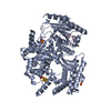
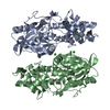
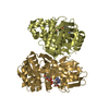
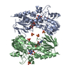
 Assembly


 Mass: 94.971 Da / Num. of mol.: 2 / Source method: obtained synthetically / Formula: PO4
 Mass: 18.015 Da / Num. of mol.: 196 / Source method: isolated from a natural source / Formula: H2O
 Sample preparation
 / Beamline: 22-ID / Wavelength: 0.9792 Å
 Processing