 Movie
Movie Controller


+ Open data

- Basic information
| Entry | Database: PDB / ID: 3nm9 | ||||||
|---|---|---|---|---|---|---|---|
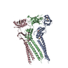
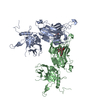
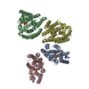
| Title | HMGD(M13A)-DNA complex | ||||||
 Components Components |
| ||||||
 Keywords Keywords | GENE REGULATION/DNA / High mobility group / DNA bending / non-sequence-specific / HMG domain / chromosomal protein / DNA / GENE REGULATION-DNA complex | ||||||
| Function / homology |  Function and homology information Function and homology informationMitochondrial transcription initiation / Mitochondrial protein degradation / DNA binding, bending / minor groove of adenine-thymine-rich DNA binding / chromatin organization / chromatin remodeling / chromatin / DNA binding / nucleus Similarity search - Function | ||||||
| Biological species |  | ||||||
| Method |  X-RAY DIFFRACTION / MOLECULAR REPLACEMENT / Resolution: 2.85 Å X-RAY DIFFRACTION / MOLECULAR REPLACEMENT / Resolution: 2.85 Å | ||||||
 Authors Authors | Churchill, M.E.A. / Klass, J. / Zoetewey, D.L. | ||||||
 Citation Citation | Journal: J.Mol.Biol. / Year: 2010 Title: Structural analysis of HMGD-DNA complexes reveals influence of intercalation on sequence selectivity and DNA bending. Authors: Churchill, M.E. / Klass, J. / Zoetewey, D.L. | ||||||
| History |
|
- Structure visualization
| Structure viewer | Molecule: MolmilJmol/JSmol |
|---|
- Downloads & links
-Download
| PDBx/mmCIF format | 3nm9.cif.gz | 150.7 KB | Display | PDBx/mmCIF format |
|---|---|---|---|---|
| PDB format | pdb3nm9.ent.gz | 116.2 KB | Display | PDB format |
| PDBx/mmJSON format | 3nm9.json.gz | Tree view | PDBx/mmJSON format | |
| Others |  Other downloads Other downloads |
-Validation report
| Arichive directory | https://data.pdbj.org/pub/pdb/validation_reports/nm/3nm9ftp://data.pdbj.org/pub/pdb/validation_reports/nm/3nm9 | HTTPS FTP |
|---|
-Related structure data
| Related structure data | 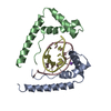 1qrvS S: Starting model for refinement |
|---|---|
| Similar structure data |

-Links
 PDBj
PDBj


- Assembly
| Deposited unit | 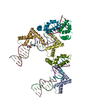
| ||||||||
|---|---|---|---|---|---|---|---|---|---|
| 1 |
| ||||||||
| Unit cell |
|
-Components
| #1: Protein | Mass: 8310.396 Da / Num. of mol.: 6 / Mutation: M13A Source method: isolated from a genetically manipulated source Source: (gene. exp.)  #2: DNA chain | Mass: 3374.210 Da / Num. of mol.: 10 / Source method: obtained synthetically #3: Water | ChemComp-HOH / |  Mass: 18.015 Da / Num. of mol.: 4 / Source method: isolated from a natural source / Formula: H2O Mass: 18.015 Da / Num. of mol.: 4 / Source method: isolated from a natural source / Formula: H2O |
|---|
-Experimental details
-Experiment
| Experiment | Method: X-RAY DIFFRACTION / Number of used crystals: 1 |
|---|
- Sample preparation
| Crystal | Density Matthews: 3.26 Å3/Da / Density % sol: 62.27 % |
|---|---|
| Crystal grow | Temperature: 297 K / Method: vapor diffusion, hanging drop / pH: 5.25 Details: 1.2 mM HMGDM13A protein, 1.18 mM duplex DNA fragment (GCGATATCGC), 5 mM MES-Na pH 5.25, 10 mM NaCl, and 7.6% PEG 3350 equilibrated against 0.5 mL 32% PEG 3350, VAPOR DIFFUSION, HANGING DROP, temperature 297K |
-Data collection
| Diffraction | Mean temperature: 100 K |
|---|---|
| Diffraction source | Source: ROTATING ANODE / Type: RIGAKU RUH3R / Wavelength: 1.5418 Å |
| Detector | Type: RIGAKU RAXIS IV++ / Detector: IMAGE PLATE / Date: Jan 1, 2001 / Details: Blue Optics |
| Radiation | Protocol: SINGLE WAVELENGTH / Monochromatic (M) / Laue (L): M / Scattering type: x-ray |
| Radiation wavelength | Wavelength: 1.5418 Å / Relative weight: 1 |
| Reflection | Resolution: 2.8→20 Å / Num. obs: 23992 / % possible obs: 92.3 % / Observed criterion σ(F): 0 / Observed criterion σ(I): 0 / Redundancy: 2.6 % / Rsym value: 0.06 / Net I/σ(I): 21.9 |
| Reflection shell | Resolution: 2.8→2.91 Å / Mean I/σ(I) obs: 4.6 / Rsym value: 0.208 / % possible all: 82.1 |
- Processing
| Software |
| ||||||||||||||||||||||||||||||||||||||||||||||||||||||||||||||||||||||||||||||||||||||||||||||||||||||||||||||||||||||||||||||||||||||||||||||||||||||||||||||||||||||||||
|---|---|---|---|---|---|---|---|---|---|---|---|---|---|---|---|---|---|---|---|---|---|---|---|---|---|---|---|---|---|---|---|---|---|---|---|---|---|---|---|---|---|---|---|---|---|---|---|---|---|---|---|---|---|---|---|---|---|---|---|---|---|---|---|---|---|---|---|---|---|---|---|---|---|---|---|---|---|---|---|---|---|---|---|---|---|---|---|---|---|---|---|---|---|---|---|---|---|---|---|---|---|---|---|---|---|---|---|---|---|---|---|---|---|---|---|---|---|---|---|---|---|---|---|---|---|---|---|---|---|---|---|---|---|---|---|---|---|---|---|---|---|---|---|---|---|---|---|---|---|---|---|---|---|---|---|---|---|---|---|---|---|---|---|---|---|---|---|---|---|---|---|
| Refinement | Method to determine structure: MOLECULAR REPLACEMENT Starting model: 1qrv Resolution: 2.85→20 Å / Cor.coef. Fo:Fc: 0.907 / Cor.coef. Fo:Fc free: 0.855 / SU B: 12.834 / SU ML: 0.251 / Cross valid method: THROUGHOUT / ESU R Free: 0.444 / Stereochemistry target values: MAXIMUM LIKELIHOOD
| ||||||||||||||||||||||||||||||||||||||||||||||||||||||||||||||||||||||||||||||||||||||||||||||||||||||||||||||||||||||||||||||||||||||||||||||||||||||||||||||||||||||||||
| Solvent computation | Ion probe radii: 0.8 Å / Shrinkage radii: 0.8 Å / VDW probe radii: 1.2 Å / Solvent model: MASK | ||||||||||||||||||||||||||||||||||||||||||||||||||||||||||||||||||||||||||||||||||||||||||||||||||||||||||||||||||||||||||||||||||||||||||||||||||||||||||||||||||||||||||
| Displacement parameters | Biso mean: 58.037 Å2
| ||||||||||||||||||||||||||||||||||||||||||||||||||||||||||||||||||||||||||||||||||||||||||||||||||||||||||||||||||||||||||||||||||||||||||||||||||||||||||||||||||||||||||
| Refinement step | Cycle: LAST / Resolution: 2.85→20 Å
| ||||||||||||||||||||||||||||||||||||||||||||||||||||||||||||||||||||||||||||||||||||||||||||||||||||||||||||||||||||||||||||||||||||||||||||||||||||||||||||||||||||||||||
| Refine LS restraints |
| ||||||||||||||||||||||||||||||||||||||||||||||||||||||||||||||||||||||||||||||||||||||||||||||||||||||||||||||||||||||||||||||||||||||||||||||||||||||||||||||||||||||||||
| LS refinement shell | Resolution: 2.85→2.923 Å / Total num. of bins used: 20
|
