Mass: 18.015 Da / Num. of mol.: 141 / Source method: isolated from a natural source / Formula: H2O
Has protein modification
Y
Sequence details
THE CONSTRUCT WAS EXPRESSED WITH AN N-TERMINAL PURIFICATION TAG MGSDKIHHHHHHENLYFQG. THE TAG WAS ...THE CONSTRUCT WAS EXPRESSED WITH AN N-TERMINAL PURIFICATION TAG MGSDKIHHHHHHENLYFQG. THE TAG WAS REMOVED WITH TEV PROTEASE LEAVING ONLY A GLYCINE (0) FOLLOWED BY THE TARGET SEQUENCE.
-
Experimental details
-
Experiment
Experiment
Method: X-RAY DIFFRACTION / Number of used crystals: 1
-
Sample preparation
Crystal
Density Matthews: 2.16 Å3/Da / Density % sol: 43.07 %
Crystal grow
Temperature: 277 K / pH: 7.5 Details: 1.4000M Na3Citrate, 0.1M HEPES pH 7.5, NANODROP, VAPOR DIFFUSION, SITTING DROP, temperature 277K
Resolution: 1.78→39.621 Å / Num. obs: 15531 / % possible obs: 98.1 % / Redundancy: 2.8 % / Biso Wilson estimate: 21.33 Å2 / Rmerge(I) obs: 0.069 / Rsym value: 0.069 / Net I/σ(I): 9.8
Reflection shell
Resolution (Å)
Rmerge(I) obs
Mean I/σ(I) obs
Num. measured obs
Num. unique obs
% possible all
2-2.05
0.943
2.4
31503
4244
99.9
2.05-2.11
0.788
2.9
30771
4135
100
2.11-2.17
0.638
3.5
29991
4033
100
2.17-2.24
0.516
4.2
29054
3909
100
2.24-2.31
0.469
4.6
28142
3791
100
2.31-2.39
0.402
5.3
27337
3685
100
2.39-2.48
0.338
6.1
26546
3685
100
2.48-2.58
0.304
6.6
25421
3574
100
2.58-2.7
0.236
8.3
24463
3432
100
2.7-2.83
0.215
8.9
23356
3303
100
2.83-2.98
0.166
10.9
22400
3157
100
2.98-3.16
0.132
13.1
21158
3043
100
3.16-3.38
0.11
15.2
19766
2688
100
3.38-3.65
0.092
18.1
18425
2516
100
3.65-4
0.077
21.7
17034
2343
100
4-4.47
0.063
24.8
15489
2147
100
4.47-5.16
0.06
24.6
13543
1894
100
5.16-6.32
0.067
20.7
11606
1650
100
6.32-8.94
0.056
23.8
8895
1300
100
8.94-29.7
0.048
24.7
4719
765
100
-
Phasing
Phasing
Method: MAD
-
Processing
Software
Name
Version
Classification
NB
REFMAC
5.5.0110
refinement
PHENIX
refinement
SHELX
phasing
MolProbity
3beta29
modelbuilding
SCALA
3.2.25
datascaling
PDB_EXTRACT
3.006
dataextraction
MOSFLM
datareduction
SHELXD
phasing
autoSHARP
phasing
Refinement
Method to determine structure: MAD / Resolution: 1.78→39.62 Å / Cor.coef. Fo:Fc: 0.966 / Cor.coef. Fo:Fc free: 0.948 / Occupancy max: 1 / Occupancy min: 0.5 / SU B: 6.454 / SU ML: 0.099 / Cross valid method: THROUGHOUT / σ(F): 0 / ESU R: 0.133 / ESU R Free: 0.129 Stereochemistry target values: MAXIMUM LIKELIHOOD WITH PHASES Details: 1. HYDROGENS HAVE BEEN ADDED IN THE RIDING POSITIONS. 2. A MET-INHIBITION PROTOCOL WAS USED FOR SELENOMETHIONINE INCORPORATION DURING PROTEIN EXPRESSION. THE OCCUPANCY OF THE SE ATOMS IN THE ...Details: 1. HYDROGENS HAVE BEEN ADDED IN THE RIDING POSITIONS. 2. A MET-INHIBITION PROTOCOL WAS USED FOR SELENOMETHIONINE INCORPORATION DURING PROTEIN EXPRESSION. THE OCCUPANCY OF THE SE ATOMS IN THE MSE RESIDUES WAS REDUCED TO 0.75 TO ACCOUNT FOR THE REDUCED SCATTERING POWER DUE TO PARTIAL S-MET INCORPORATION. 3. ATOM RECORD CONTAINS SUM OF TLS AND RESIDUAL B FACTORS. ANISOU RECORD CONTAINS SUM OF TLS AND RESIDUAL U FACTORS. 4. WATERS WERE EXCLUDED FROM AUTOMATIC TLS ASSIGNMENT. 5. 1,2-ETHANEDIOL (EDO) MOLECULES FROM THE CRYOPROTECTION SOLUTION WERE MODELED INTO THE STRUCTURE.
Rfactor
Num. reflection
% reflection
Selection details
Rfree
0.222
788
5.1 %
RANDOM
Rwork
0.177
-
-
-
obs
0.179
15531
97.8 %
-
Solvent computation
Ion probe radii: 0.8 Å / Shrinkage radii: 0.8 Å / VDW probe radii: 1.4 Å / Solvent model: MASK
Displacement parameters
Biso mean: 26.66 Å2
Baniso -1
Baniso -2
Baniso -3
1-
2.57 Å2
0 Å2
0.82 Å2
2-
-
-0.84 Å2
0 Å2
3-
-
-
-1.12 Å2
Refinement step
Cycle: LAST / Resolution: 1.78→39.62 Å
Protein
Nucleic acid
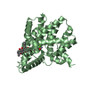
Ligand
Solvent
Total
Num. atoms
1321
0
8
141
1470
Refine LS restraints
Refine-ID
Type
Dev ideal
Dev ideal target
Number
X-RAY DIFFRACTION
r_bond_refined_d
0.015
0.022
1381
X-RAY DIFFRACTION
r_bond_other_d
0.001
0.02
943
X-RAY DIFFRACTION
r_angle_refined_deg
1.298
1.964
1875
X-RAY DIFFRACTION
r_angle_other_deg
0.85
3
2311
X-RAY DIFFRACTION
r_dihedral_angle_1_deg
4.881
5
172
X-RAY DIFFRACTION
r_dihedral_angle_2_deg
37.624
25.303
66
X-RAY DIFFRACTION
r_dihedral_angle_3_deg
12.438
15
227
X-RAY DIFFRACTION
r_dihedral_angle_4_deg
22.22
15
6
X-RAY DIFFRACTION
r_chiral_restr
0.08
0.2
201
X-RAY DIFFRACTION
r_gen_planes_refined
0.006
0.021
1554
X-RAY DIFFRACTION
r_gen_planes_other
0.001
0.02
262
X-RAY DIFFRACTION
r_nbd_refined
X-RAY DIFFRACTION
r_nbd_other
X-RAY DIFFRACTION
r_nbtor_refined
X-RAY DIFFRACTION
r_nbtor_other
X-RAY DIFFRACTION
r_xyhbond_nbd_refined
X-RAY DIFFRACTION
r_xyhbond_nbd_other
X-RAY DIFFRACTION
r_metal_ion_refined
X-RAY DIFFRACTION
r_metal_ion_other
X-RAY DIFFRACTION
r_symmetry_vdw_refined
X-RAY DIFFRACTION
r_symmetry_vdw_other
X-RAY DIFFRACTION
r_symmetry_hbond_refined
X-RAY DIFFRACTION
r_symmetry_hbond_other
X-RAY DIFFRACTION
r_symmetry_metal_ion_refined
X-RAY DIFFRACTION
r_symmetry_metal_ion_other
X-RAY DIFFRACTION
r_mcbond_it
1.768
3
859
X-RAY DIFFRACTION
r_mcbond_other
0.485
3
339
X-RAY DIFFRACTION
r_mcangle_it
2.904
5
1387
X-RAY DIFFRACTION
r_scbond_it
5.037
8
522
X-RAY DIFFRACTION
r_scangle_it
7.658
11
488
X-RAY DIFFRACTION
r_rigid_bond_restr
X-RAY DIFFRACTION
r_sphericity_free
X-RAY DIFFRACTION
r_sphericity_bonded
LS refinement shell
Resolution: 1.78→1.83 Å / Total num. of bins used: 20
Rfactor
Num. reflection
% reflection
Rfree
0.378
47
-
Rwork
0.354
1082
-
obs
-
-
97.58 %
Refinement TLS params.
Method: refined / Origin x: -4.05 Å / Origin y: 17.88 Å / Origin z: 19.972 Å
11
12
13
21
22
23
31
32
33
T
0.1407 Å2
-0.0075 Å2
0.0422 Å2
-
0.0096 Å2
0.0022 Å2
-
-
0.0341 Å2
L
0.672 °2
-0.0332 °2
-0.4488 °2
-
0.8813 °2
0.1025 °2
-
-
1.1449 °2
S
0.0105 Å °
-0.0248 Å °
-0.0082 Å °
0.01 Å °
0.0255 Å °
-0.0488 Å °
-0.0634 Å °
0.0751 Å °
-0.0361 Å °
+
About Yorodumi
-
News
-
Feb 9, 2022. New format data for meta-information of EMDB entries
New format data for meta-information of EMDB entries
Version 3 of the EMDB header file is now the official format.
The previous official version 1.9 will be removed from the archive.
In the structure databanks used in Yorodumi, some data are registered as the other names, "COVID-19 virus" and "2019-nCoV". Here are the details of the virus and the list of structure data.
Jan 31, 2019. EMDB accession codes are about to change! (news from PDBe EMDB page)
EMDB accession codes are about to change! (news from PDBe EMDB page)
The allocation of 4 digits for EMDB accession codes will soon come to an end. Whilst these codes will remain in use, new EMDB accession codes will include an additional digit and will expand incrementally as the available range of codes is exhausted. The current 4-digit format prefixed with “EMD-” (i.e. EMD-XXXX) will advance to a 5-digit format (i.e. EMD-XXXXX), and so on. It is currently estimated that the 4-digit codes will be depleted around Spring 2019, at which point the 5-digit format will come into force.
The EM Navigator/Yorodumi systems omit the EMD- prefix.
Related info.:Q: What is EMD? / ID/Accession-code notation in Yorodumi/EM Navigator
Yorodumi is a browser for structure data from EMDB, PDB, SASBDB, etc.
This page is also the successor to EM Navigator detail page, and also detail information page/front-end page for Omokage search.
The word "yorodu" (or yorozu) is an old Japanese word meaning "ten thousand". "mi" (miru) is to see.
Related info.:EMDB / PDB / SASBDB / Comparison of 3 databanks / Yorodumi Search / Aug 31, 2016. New EM Navigator & Yorodumi / Yorodumi Papers / Jmol/JSmol / Function and homology information / Changes in new EM Navigator and Yorodumi
 Movie
Movie Controller

 Yorodumi
 Open data
 Basic information
 Components
 Keywords
 Function and homology information
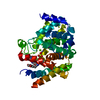
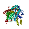
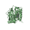
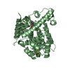
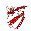
 Exiguobacterium sibiricum 255-15 (bacteria)
 X-RAY DIFFRACTION /
  Authors
 Citation
 Structure visualization
 Downloads & links
 Other downloads


 PDBj
PDBj Assembly
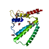

 Mass: 62.068 Da / Num. of mol.: 2 / Source method: obtained synthetically / Formula: C2H6O2
 Mass: 18.015 Da / Num. of mol.: 141 / Source method: isolated from a natural source / Formula: H2O
 Sample preparation
 / Beamline: 8.2.2 / Wavelength: 1.0000,0.9798
 Processing