 Movie
Movie Controller

[English] 日本語
 Yorodumi
- PDB-3nkt: Crystal Structure of Salicylate 1,2-dioxygenase from Pseudoaminob... -
+ Open data

- Basic information
| Entry | Database: PDB / ID: 3nkt | ||||||
|---|---|---|---|---|---|---|---|
| Title | Crystal Structure of Salicylate 1,2-dioxygenase from Pseudoaminobacter salicylatoxidans Adducts with naphthoate | ||||||
 Components Components | Gentisate 1,2-Dioxygenase | ||||||
 Keywords Keywords | OXIDOREDUCTASE / BETA-SANDWICH | ||||||
| Function / homology |  Function and homology information Function and homology information | ||||||
| Biological species |  Pseudaminobacter salicylatoxidans (bacteria) Pseudaminobacter salicylatoxidans (bacteria) | ||||||
| Method |  X-RAY DIFFRACTION / SYNCHROTRON / FOURIER SYNTHESIS / Resolution: 2.35 Å X-RAY DIFFRACTION / SYNCHROTRON / FOURIER SYNTHESIS / Resolution: 2.35 Å | ||||||
 Authors Authors | Ferraroni, M. / Briganti, F. / Matera, I. | ||||||
 Citation Citation | Journal: J.Struct.Biol. / Year: 2012 Title: Crystal structures of salicylate 1,2-dioxygenase-substrates adducts: A step towards the comprehension of the structural basis for substrate selection in class III ring cleaving dioxygenases. Authors: Ferraroni, M. / Matera, I. / Steimer, L. / Burger, S. / Scozzafava, A. / Stolz, A. / Briganti, F. | ||||||
| History |
|
- Structure visualization
| Structure viewer | Molecule: MolmilJmol/JSmol |
|---|
- Downloads & links
-Download
| PDBx/mmCIF format | 3nkt.cif.gz | 92.2 KB | Display | PDBx/mmCIF format |
|---|---|---|---|---|
| PDB format | pdb3nkt.ent.gz | 68.8 KB | Display | PDB format |
| PDBx/mmJSON format | 3nkt.json.gz | Tree view | PDBx/mmJSON format | |
| Others |  Other downloads Other downloads |
-Validation report
| Arichive directory | https://data.pdbj.org/pub/pdb/validation_reports/nk/3nktftp://data.pdbj.org/pub/pdb/validation_reports/nk/3nkt | HTTPS FTP |
|---|
-Related structure data


-Links
 PDBj
PDBj


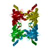
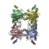
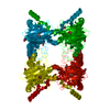
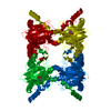
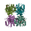
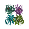
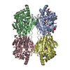
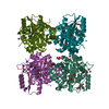
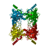
- Assembly
| Deposited unit | 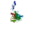
| ||||||||
|---|---|---|---|---|---|---|---|---|---|
| 1 | 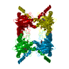
| ||||||||
| Unit cell |
|
-Components
| #1: Protein | Mass: 41144.438 Da / Num. of mol.: 1 Source method: isolated from a genetically manipulated source Source: (gene. exp.) Pseudaminobacter salicylatoxidans (bacteria)Strain: BN12 / Plasmid: pET28A / Production host: | ||
|---|---|---|---|
| #2: Chemical | ChemComp-FE2 /   Mass: 55.845 Da / Num. of mol.: 1 / Source method: obtained synthetically / Formula: Fe Mass: 55.845 Da / Num. of mol.: 1 / Source method: obtained synthetically / Formula: Fe | ||
| #3: Chemical | ChemComp-1HN /   Mass: 188.179 Da / Num. of mol.: 1 / Source method: obtained synthetically / Formula: C11H8O3 Mass: 188.179 Da / Num. of mol.: 1 / Source method: obtained synthetically / Formula: C11H8O3 | ||
| #4: Chemical |   Mass: 92.094 Da / Num. of mol.: 2 / Source method: obtained synthetically / Formula: C3H8O3 Mass: 92.094 Da / Num. of mol.: 2 / Source method: obtained synthetically / Formula: C3H8O3#5: Water | ChemComp-HOH / |  Mass: 18.015 Da / Num. of mol.: 308 / Source method: isolated from a natural source / Formula: H2O Mass: 18.015 Da / Num. of mol.: 308 / Source method: isolated from a natural source / Formula: H2O |
-Experimental details
-Experiment
| Experiment | Method: X-RAY DIFFRACTION / Number of used crystals: 1 |
|---|
- Sample preparation
| Crystal | Density Matthews: 3.33 Å3/Da / Density % sol: 63.02 % |
|---|---|
| Crystal grow | Temperature: 277 K / Method: vapor diffusion / pH: 8 Details: 8% PEG10000, pH 8.0, vapor diffusion, temperature 277K |
-Data collection
| Diffraction | Mean temperature: 100 K |
|---|---|
| Diffraction source | Source: SYNCHROTRON / Site: EMBL/DESY, HAMBURG  / Beamline: X13 / Wavelength: 0.8123 Å / Beamline: X13 / Wavelength: 0.8123 Å |
| Detector | Type: MAR CCD 165 mm / Detector: CCD / Date: Jan 1, 2009 |
| Radiation | Monochromator: Si (111), horizontally focusing / Protocol: SINGLE WAVELENGTH / Monochromatic (M) / Laue (L): M / Scattering type: x-ray |
| Radiation wavelength | Wavelength: 0.8123 Å / Relative weight: 1 |
| Reflection | Resolution: 2.35→46.7 Å / Num. all: 21895 / Num. obs: 21895 / % possible obs: 94.7 % / Redundancy: 4.6 % / Rmerge(I) obs: 0.115 / Rsym value: 0.115 / Net I/σ(I): 11.2 |
| Reflection shell | Resolution: 2.35→2.48 Å / Redundancy: 4.7 % / Rmerge(I) obs: 0.44 / Mean I/σ(I) obs: 2.47 / Rsym value: 0.44 / % possible all: 96 |
- Processing
| Software |
| |||||||||||||||||||||||||||||||||||||||||||||||||||||||||||||||||
|---|---|---|---|---|---|---|---|---|---|---|---|---|---|---|---|---|---|---|---|---|---|---|---|---|---|---|---|---|---|---|---|---|---|---|---|---|---|---|---|---|---|---|---|---|---|---|---|---|---|---|---|---|---|---|---|---|---|---|---|---|---|---|---|---|---|---|
| Refinement | Method to determine structure: FOURIER SYNTHESIS / Resolution: 2.35→30 Å / Cor.coef. Fo:Fc: 0.953 / Cor.coef. Fo:Fc free: 0.918 / WRfactor Rfree: 0.201 / WRfactor Rwork: 0.1531 / Occupancy max: 1 / Occupancy min: 0.5 / FOM work R set: 0.8471 / SU B: 6.259 / SU ML: 0.15 / SU R Cruickshank DPI: 0.2667 / SU Rfree: 0.2232 / Isotropic thermal model: isotropic / Cross valid method: THROUGHOUT / σ(F): 0 / ESU R Free: 0.223 / Stereochemistry target values: MAXIMUM LIKELIHOOD
| |||||||||||||||||||||||||||||||||||||||||||||||||||||||||||||||||
| Solvent computation | Ion probe radii: 0.8 Å / Shrinkage radii: 0.8 Å / VDW probe radii: 1.4 Å / Solvent model: BABINET MODEL WITH MASK | |||||||||||||||||||||||||||||||||||||||||||||||||||||||||||||||||
| Displacement parameters | Biso max: 74.22 Å2 / Biso mean: 38.9106 Å2 / Biso min: 11.59 Å2
| |||||||||||||||||||||||||||||||||||||||||||||||||||||||||||||||||
| Refinement step | Cycle: LAST / Resolution: 2.35→30 Å
| |||||||||||||||||||||||||||||||||||||||||||||||||||||||||||||||||
| Refine LS restraints |
| |||||||||||||||||||||||||||||||||||||||||||||||||||||||||||||||||
| LS refinement shell | Resolution: 2.35→2.411 Å / Total num. of bins used: 20
|
