 Movie
Movie Controller


+ Open data

- Basic information
| Entry | Database: PDB / ID: 3njw | ||||||
|---|---|---|---|---|---|---|---|
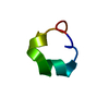
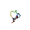
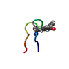
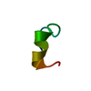
| Title | First High Resolution Crystal Structure of a Lasso Peptide | ||||||
 Components Components | Bicyclic peptide BI-32169 | ||||||
 Keywords Keywords | ANTIMICROBIAL PROTEIN / lasso peptide / lariat / protoknot / BI-32169 / glucagon receptor antagonist / microbial peptide | ||||||
| Biological species |  Streptomyces sp. (bacteria) Streptomyces sp. (bacteria) | ||||||
| Method |  X-RAY DIFFRACTION / AB INITIO / Resolution: 0.86 Å X-RAY DIFFRACTION / AB INITIO / Resolution: 0.86 Å | ||||||
 Authors Authors | Nar, H. / Schmid, A. / Puder, C. / Potterat, O. | ||||||
 Citation Citation | Journal: Chemmedchem / Year: 2010 Title: High-resolution crystal structure of a lasso Peptide. Authors: Nar, H. / Schmid, A. / Puder, C. / Potterat, O. | ||||||
| History |
|
- Structure visualization
| Structure viewer | Molecule:  MolmilJmol/JSmol MolmilJmol/JSmol |
|---|
- Downloads & links
-Download
| PDBx/mmCIF format | 3njw.cif.gz | 17 KB | Display | PDBx/mmCIF format |
|---|---|---|---|---|
| PDB format | pdb3njw.ent.gz | 10.1 KB | Display | PDB format |
| PDBx/mmJSON format | 3njw.json.gz | Tree view | PDBx/mmJSON format | |
| Others |  Other downloads Other downloads |
-Validation report
| Summary document | 3njw_validation.pdf.gz | 383.5 KB | Display | wwPDB validaton report |
|---|---|---|---|---|
| Full document | 3njw_full_validation.pdf.gz | 383.5 KB | Display | |
| Data in XML | 3njw_validation.xml.gz | 3.3 KB | Display | |
| Data in CIF | 3njw_validation.cif.gz | 3.8 KB | Display | |
| Arichive directory | https://data.pdbj.org/pub/pdb/validation_reports/nj/3njwftp://data.pdbj.org/pub/pdb/validation_reports/nj/3njw | HTTPS FTP |
-Related structure data
| Similar structure data |
|---|


-Links
 PDBj
PDBj- Assembly
| Deposited unit | 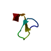
| ||||||||
|---|---|---|---|---|---|---|---|---|---|
| 1 |
| ||||||||
| Unit cell |
|
-Components
| #1: Protein/peptide | Mass: 2057.310 Da / Num. of mol.: 1 Source method: isolated from a genetically manipulated source Source: (gene. exp.) Streptomyces sp. (bacteria) / Strain: DSM 14996 / Production host: Streptomyces sp. (bacteria) |
|---|---|
| #2: Water | ChemComp-HOH /  Mass: 18.015 Da / Num. of mol.: 25 / Source method: isolated from a natural source / Formula: H2O Mass: 18.015 Da / Num. of mol.: 25 / Source method: isolated from a natural source / Formula: H2O |
| Has protein modification | Y |
-Experimental details
-Experiment
| Experiment | Method: X-RAY DIFFRACTION / Number of used crystals: 1 |
|---|
- Sample preparation
| Crystal | Density Matthews: 1.5 Å3/Da / Density % sol: 17.82 % |
|---|---|
| Crystal grow | Temperature: 293 K / Method: vapor diffusion, hanging drop / pH: 8.5 Details: BI-32169 peptide in 50% DMSO/water mixture equilibrated against 20-50% MPD, 0.1-0.5M sodium phosphate or ammonium sulphate, 0.1M Tris HCl, pH 8.5, VAPOR DIFFUSION, HANGING DROP, temperature 293K |
-Data collection
| Diffraction | Mean temperature: 100 K |
|---|---|
| Diffraction source | Source: ROTATING ANODE / Type: RIGAKU RU300 / Wavelength: 1.54 Å |
| Detector | Type: RIGAKU SATURN 944 / Detector: CCD / Date: Oct 25, 2006 / Details: multilayer optics |
| Radiation | Monochromator: multilayer optics / Protocol: SINGLE WAVELENGTH / Monochromatic (M) / Laue (L): M / Scattering type: x-ray |
| Radiation wavelength | Wavelength: 1.54 Å / Relative weight: 1 |
| Reflection | Resolution: 0.86→8.94 Å / Num. all: 19700 / Num. obs: 19602 / % possible obs: 98.7 % / Redundancy: 7.1 % / Rmerge(I) obs: 0.076 / Net I/σ(I): 16 |
| Reflection shell | Resolution: 0.86→0.9 Å / Rmerge(I) obs: 0.41 / Mean I/σ(I) obs: 2.1 / % possible all: 87.2 |
- Processing
| Software |
| ||||||||||||||||||||
|---|---|---|---|---|---|---|---|---|---|---|---|---|---|---|---|---|---|---|---|---|---|
| Refinement | Method to determine structure: AB INITIO / Resolution: 0.86→8.94 Å / Num. parameters: 1443 / Num. restraintsaints: 0 / Cross valid method: THROUGHOUT / σ(F): 4
| ||||||||||||||||||||
| Refine analyze | Num. disordered residues: 0 / Occupancy sum hydrogen: 124 / Occupancy sum non hydrogen: 161 | ||||||||||||||||||||
| Refinement step | Cycle: LAST / Resolution: 0.86→8.94 Å
| ||||||||||||||||||||
| LS refinement shell | Highest resolution: 0.86 Å / Num. reflection obs: 16628 |
