 Movie
Movie Controller

[English] 日本語
 Yorodumi
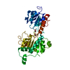
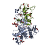
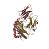
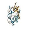
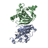
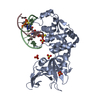
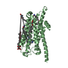
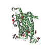
- PDB-3nj2: Crystal structure of cce_0566 from the cyanobacterium Cyanothece ... -
+ Open data

- Basic information
| Entry | Database: PDB / ID: 3nj2 | ||||||
|---|---|---|---|---|---|---|---|
| Title | Crystal structure of cce_0566 from the cyanobacterium Cyanothece 51142, a protein associated with nitrogen fixation from the DUF269 family | ||||||
 Components Components | DUF269-containing protein | ||||||
 Keywords Keywords | UNKNOWN FUNCTION / cyanobacteria / circadium rhythms / nitrogen fixation | ||||||
| Function / homology | Protein of unknown function DUF269 / Uncharacterised protein family UPF0460 / Protein of unknown function, DUF269 / Putative cytoplasmic protein / Orthogonal Bundle / Mainly Alpha / DUF269-containing protein Function and homology information Function and homology information | ||||||
| Biological species |  Cyanothece sp. ATCC 51142 (bacteria) Cyanothece sp. ATCC 51142 (bacteria) | ||||||
| Method |  X-RAY DIFFRACTION / SYNCHROTRON / MOLECULAR REPLACEMENT / Resolution: 1.59 Å X-RAY DIFFRACTION / SYNCHROTRON / MOLECULAR REPLACEMENT / Resolution: 1.59 Å | ||||||
 Authors Authors | Robinson, H. / Ralston, C.Y. / Addlagatta, A. / Buchko, G.W. | ||||||
 Citation Citation | Journal: Febs Lett. / Year: 2012 Title: Crystal structure of cce_0566 from Cyanothece 51142, a protein associated with nitrogen fixation in the DUF269 family. Authors: Buchko, G.W. / Robinson, H. | ||||||
| History |
|
- Structure visualization
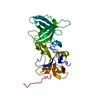
| Structure viewer | Molecule: MolmilJmol/JSmol |
|---|
- Downloads & links
-Download
| PDBx/mmCIF format | 3nj2.cif.gz | 80.2 KB | Display | PDBx/mmCIF format |
|---|---|---|---|---|
| PDB format | pdb3nj2.ent.gz | 59.6 KB | Display | PDB format |
| PDBx/mmJSON format | 3nj2.json.gz | Tree view | PDBx/mmJSON format | |
| Others |  Other downloads Other downloads |
-Validation report
| Arichive directory | https://data.pdbj.org/pub/pdb/validation_reports/nj/3nj2ftp://data.pdbj.org/pub/pdb/validation_reports/nj/3nj2 | HTTPS FTP |
|---|
-Related structure data
| Related structure data | 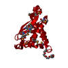 3g7pS S: Starting model for refinement |
|---|---|
| Similar structure data |

-Links
 PDBj
PDBj- Assembly
| Deposited unit | 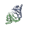
| ||||||||
|---|---|---|---|---|---|---|---|---|---|
| 1 |
| ||||||||
| Unit cell |
|
-Components
| #1: Protein | Mass: 19729.605 Da / Num. of mol.: 2 / Fragment: UNP residues 19-171 Source method: isolated from a genetically manipulated source Details: NdeI, BamHI site / Source: (gene. exp.) Cyanothece sp. ATCC 51142 (bacteria) / Gene: cce_0566 / Plasmid: pET28b / Production host: #2: Water | ChemComp-HOH / |  Mass: 18.015 Da / Num. of mol.: 300 / Source method: isolated from a natural source / Formula: H2O Mass: 18.015 Da / Num. of mol.: 300 / Source method: isolated from a natural source / Formula: H2O |
|---|
-Experimental details
-Experiment
| Experiment | Method: X-RAY DIFFRACTION / Number of used crystals: 1 |
|---|
- Sample preparation
| Crystal | Density Matthews: 2.11 Å3/Da / Density % sol: 41.69 % |
|---|---|
| Crystal grow | Temperature: 293 K / Method: vapor diffusion, hanging drop / pH: 7.1 Details: 0.2 M Ammonium phosphate monobasic, 0.1 M Tris pH 8.5, 50% v/v (+/-)-2-Methyl-2,4-pentanediol, protein in 100 mM NaCl, 20 mM Tris, 1 mM DTT, pH 7.1. Protein 2 mg/mL, Mix 1.5 uL of protein ...Details: 0.2 M Ammonium phosphate monobasic, 0.1 M Tris pH 8.5, 50% v/v (+/-)-2-Methyl-2,4-pentanediol, protein in 100 mM NaCl, 20 mM Tris, 1 mM DTT, pH 7.1. Protein 2 mg/mL, Mix 1.5 uL of protein and precipatant, VAPOR DIFFUSION, HANGING DROP, temperature 293K |
-Data collection
| Diffraction | Mean temperature: 100 K |
|---|---|
| Diffraction source | Source: SYNCHROTRON / Site: NSLS  / Beamline: X29A / Wavelength: 0.9792 Å / Beamline: X29A / Wavelength: 0.9792 Å |
| Detector | Type: ADSC QUANTUM 315 / Detector: CCD / Date: Oct 20, 2008 |
| Radiation | Monochromator: SI(111) / Protocol: SINGLE WAVELENGTH / Monochromatic (M) / Laue (L): M / Scattering type: x-ray |
| Radiation wavelength | Wavelength: 0.9792 Å / Relative weight: 1 |
| Reflection | Resolution: 1.59→33 Å / Num. all: 38536 / Num. obs: 38536 / % possible obs: 91.692 % / Observed criterion σ(F): 0 / Observed criterion σ(I): 0 / Biso Wilson estimate: 19.938 Å2 |
| Reflection shell | Resolution: 1.593→1.634 Å / Num. unique all: 2312 / % possible all: 75.34 |
- Processing
| Software |
| ||||||||||||||||||||||||||||||||||||||||||||||||||||||||||||||||||||||||||||||||||||||||||||||||||||||||||||||||||||||||||||||||||||||||||||||||||||||||||||||||||||||||||
|---|---|---|---|---|---|---|---|---|---|---|---|---|---|---|---|---|---|---|---|---|---|---|---|---|---|---|---|---|---|---|---|---|---|---|---|---|---|---|---|---|---|---|---|---|---|---|---|---|---|---|---|---|---|---|---|---|---|---|---|---|---|---|---|---|---|---|---|---|---|---|---|---|---|---|---|---|---|---|---|---|---|---|---|---|---|---|---|---|---|---|---|---|---|---|---|---|---|---|---|---|---|---|---|---|---|---|---|---|---|---|---|---|---|---|---|---|---|---|---|---|---|---|---|---|---|---|---|---|---|---|---|---|---|---|---|---|---|---|---|---|---|---|---|---|---|---|---|---|---|---|---|---|---|---|---|---|---|---|---|---|---|---|---|---|---|---|---|---|---|---|---|
| Refinement | Method to determine structure: MOLECULAR REPLACEMENT Starting model: pdb entry 3G7P Resolution: 1.59→33 Å / Cor.coef. Fo:Fc: 0.954 / Cor.coef. Fo:Fc free: 0.928 / SU B: 1.926 / SU ML: 0.07 / Cross valid method: THROUGHOUT / σ(F): 0 / ESU R Free: 0.113 / Stereochemistry target values: MAXIMUM LIKELIHOOD / Details: HYDROGENS HAVE BEEN ADDED IN THE RIDING POSITIONS
| ||||||||||||||||||||||||||||||||||||||||||||||||||||||||||||||||||||||||||||||||||||||||||||||||||||||||||||||||||||||||||||||||||||||||||||||||||||||||||||||||||||||||||
| Solvent computation | Ion probe radii: 0.8 Å / Shrinkage radii: 0.8 Å / VDW probe radii: 1.4 Å / Solvent model: MASK | ||||||||||||||||||||||||||||||||||||||||||||||||||||||||||||||||||||||||||||||||||||||||||||||||||||||||||||||||||||||||||||||||||||||||||||||||||||||||||||||||||||||||||
| Displacement parameters | Biso mean: 21.764 Å2
| ||||||||||||||||||||||||||||||||||||||||||||||||||||||||||||||||||||||||||||||||||||||||||||||||||||||||||||||||||||||||||||||||||||||||||||||||||||||||||||||||||||||||||
| Refinement step | Cycle: LAST / Resolution: 1.59→33 Å
| ||||||||||||||||||||||||||||||||||||||||||||||||||||||||||||||||||||||||||||||||||||||||||||||||||||||||||||||||||||||||||||||||||||||||||||||||||||||||||||||||||||||||||
| Refine LS restraints |
| ||||||||||||||||||||||||||||||||||||||||||||||||||||||||||||||||||||||||||||||||||||||||||||||||||||||||||||||||||||||||||||||||||||||||||||||||||||||||||||||||||||||||||
| LS refinement shell | Resolution: 1.59→1.634 Å / Total num. of bins used: 20
|
