[English] 日本語
Yorodumi
- PDB-3ndz: The structure of the catalytic and carbohydrate binding domain of... -

+
Open data


ID or keywords:

Loading...

-
Basic information

Entry
Database: PDB / ID: 3ndz
TitleThe structure of the catalytic and carbohydrate binding domain of endoglucanase D from Clostridium cellulovorans bound to cellotriose
Components(Endoglucanase D) x 2
KeywordsHYDROLASE / cellotriose / endoglucanase / xylanase / carbohydrate binding domain / glucanase
Function / homology
Function and homology information


cellulase / polysaccharide binding / cellulase activity / cellulose catabolic process
Similarity search - Function
Immunoglobulin-like - #290 / Cellulose binding domain / Carbohydrate-binding type-2 domain / CBM2 (Carbohydrate-binding type-2) domain profile. / CBD_II / CBM2, carbohydrate-binding domain superfamily / Glycoside hydrolase, family 5, conserved site / Glycosyl hydrolases family 5 signature. / CBM2/CBM3, carbohydrate-binding domain superfamily / Glycoside hydrolase, family 5 ...Immunoglobulin-like - #290 / Cellulose binding domain / Carbohydrate-binding type-2 domain / CBM2 (Carbohydrate-binding type-2) domain profile. / CBD_II / CBM2, carbohydrate-binding domain superfamily / Glycoside hydrolase, family 5, conserved site / Glycosyl hydrolases family 5 signature. / CBM2/CBM3, carbohydrate-binding domain superfamily / Glycoside hydrolase, family 5 / Cellulase (glycosyl hydrolase family 5) / Glycosidases / Glycoside hydrolase superfamily / TIM Barrel / Alpha-Beta Barrel / Immunoglobulin-like / Sandwich / Mainly Beta / Alpha Beta
Similarity search - Domain/homology
beta-cellotriose / Endoglucanase D
Similarity search - Component
Biological speciesClostridium cellulovorans (bacteria)
MethodX-RAY DIFFRACTION / SYNCHROTRON / MOLECULAR REPLACEMENT / molecular replacement / Resolution: 2.08 Å
AuthorsBianchetti, C.M. / Smith, R.W. / Bingman, C.A. / Phillips Jr., G.N.
CitationJournal: To be Published
Title: The structure of the catalytic and carbohydrate binding domain of endoglucanase D bound to cellotriose
Authors: Bianchetti, C.M. / Smith, R.W. / Bingman, C.A. / Phillips Jr., G.N.
History
DepositionJun 8, 2010Deposition site: RCSB / Processing site: RCSB
Revision 1.0Jun 29, 2011Provider: repository / Type: Initial release
Revision 1.1Jul 13, 2011Group: Version format compliance
Revision 2.0Jul 29, 2020Group: Atomic model / Data collection ...Atomic model / Data collection / Derived calculations / Non-polymer description / Structure summary
Category: atom_site / chem_comp ...atom_site / chem_comp / entity / entity_name_com / pdbx_branch_scheme / pdbx_chem_comp_identifier / pdbx_entity_branch / pdbx_entity_branch_descriptor / pdbx_entity_branch_link / pdbx_entity_branch_list / pdbx_entity_nonpoly / pdbx_molecule_features / pdbx_nonpoly_scheme / struct_conn / struct_site / struct_site_gen
Item: _atom_site.B_iso_or_equiv / _atom_site.Cartn_x ..._atom_site.B_iso_or_equiv / _atom_site.Cartn_x / _atom_site.Cartn_y / _atom_site.Cartn_z / _atom_site.auth_asym_id / _atom_site.auth_atom_id / _atom_site.auth_comp_id / _atom_site.auth_seq_id / _atom_site.label_atom_id / _atom_site.label_comp_id / _chem_comp.formula / _chem_comp.formula_weight / _chem_comp.id / _chem_comp.name / _chem_comp.pdbx_synonyms / _chem_comp.type / _entity.formula_weight / _entity.pdbx_description / _entity.src_method / _entity.type
Description: Carbohydrate remediation / Provider: repository / Type: Remediation
Revision 2.1Feb 21, 2024Group: Data collection / Database references / Structure summary
Category: chem_comp / chem_comp_atom ...chem_comp / chem_comp_atom / chem_comp_bond / database_2
Item: _chem_comp.pdbx_synonyms / _database_2.pdbx_DOI / _database_2.pdbx_database_accession

-
Structure visualization

Structure viewerMolecule:
MolmilJmol/JSmol

Downloads & links

-
Assembly

Deposited unit
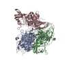
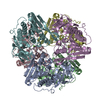
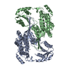
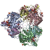
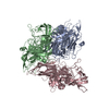
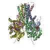
A: Endoglucanase D
E: Endoglucanase D
B: Endoglucanase D
F: Endoglucanase D
C: Endoglucanase D
G: Endoglucanase D
D: Endoglucanase D
H: Endoglucanase D
hetero molecules


Theoretical massNumber of molelcules
Total (without water)201,35614
Polymers198,3298
Non-polymers3,0276
Water34,7151927
1


  • Idetical with deposited unit
  • defined by author&software
TypeNameSymmetry operationNumber
identity operation1_555x,y,z1
Buried area19760 Å2
ΔGint-86 kcal/mol
Surface area58770 Å2
MethodPISA
Unit cell
Length a, b, c (Å)85.297, 119.050, 198.487
Angle α, β, γ (deg.)90.00, 90.00, 90.00
Int Tables number19
Space group name H-MP212121

-
Components

#1: Protein
Endoglucanase D / Endo-1 / 4-beta-glucanase D / Cellulase D


Mass: 38458.098 Da / Num. of mol.: 4 / Fragment: UNP residues 32-376
Source method: isolated from a genetically manipulated source
Source: (gene. exp.) Clostridium cellulovorans (bacteria) / Gene: engD / Plasmid: pBAD/thio-TOPO / Production host: Escherichia coli (E. coli) / Strain (production host): TOP10 / References: UniProt: P28623, cellulase
#2: Protein
Endoglucanase D / Endo-1 / 4-beta-glucanase D / Cellulase D


Mass: 11124.271 Da / Num. of mol.: 4 / Fragment: UNP residues 409-515
Source method: isolated from a genetically manipulated source
Source: (gene. exp.) Clostridium cellulovorans (bacteria) / Gene: engD / Plasmid: pBAD/thio-TOPO / Production host: Escherichia coli (E. coli) / Strain (production host): TOP10 / References: UniProt: P28623, cellulase
#3: Polysaccharide
beta-D-glucopyranose-(1-4)-beta-D-glucopyranose-(1-4)-beta-D-glucopyranose / beta-cellotriose


Type: oligosaccharide, Oligosaccharide / Class: Metabolism / Mass: 504.438 Da / Num. of mol.: 6
Source method: isolated from a genetically manipulated source
Details: oligosaccharide / References: beta-cellotriose
DescriptorTypeProgram
DGlcpb1-4DGlcpb1-4DGlcpb1-ROHGlycam Condensed SequenceGMML 1.0
WURCS=2.0/1,3,2/[a2122h-1b_1-5]/1-1-1/a4-b1_b4-c1WURCSPDB2Glycan 1.1.0
[][b-D-Glcp]{[(4+1)][b-D-Glcp]{[(4+1)][b-D-Glcp]{}}}LINUCSPDB-CARE
#4: Water ChemComp-HOH / water


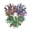
Mass: 18.015 Da / Num. of mol.: 1927 / Source method: isolated from a natural source / Formula: H2O
Compound detailsAUTHORS CRYSTALLIZED THE SEQUENCE CORRESPONDING TO UNP RESIDUES 32-515. HOWEVER, THIS ENTRY WAS ...AUTHORS CRYSTALLIZED THE SEQUENCE CORRESPONDING TO UNP RESIDUES 32-515. HOWEVER, THIS ENTRY WAS SPILT INTO 2 MOLECULES, (CHAINS A,B,C,D) AND (CHAINS E,F,G,H), SINCE THE FLEXIBLE LINKER (RESIDUES 377-408), JOINING THE N-TERMINAL DOMAIN TO THE C-TERMINAL DOMAIN WAS NOT OBSERVED IN THE ELECTRON DENSITY MAP. THUS, IT IS IMPOSSIBLE TO KNOW FOR SURE WHICH N-TERMINAL DOMAIN (A,B,C,D) IS ASSOCIATED WITH WHICH C-TERMINAL DOMAIN (E,F,G,H) IN THIS CASE.
Sequence detailsRESIDUES 'TTTPTTPTTPTTPTTPTTPTTPTTPTTPTTPQ' CONSTITUTE THE FLEXIBLE LINKER THAT WAS NOT OBSERVED IN ...RESIDUES 'TTTPTTPTTPTTPTTPTTPTTPTTPTTPTTPQ' CONSTITUTE THE FLEXIBLE LINKER THAT WAS NOT OBSERVED IN THE ELECTRON DENSITY MAP

-
Experimental details

-
Experiment

ExperimentMethod: X-RAY DIFFRACTION / Number of used crystals: 1

-
Sample preparation

CrystalDensity Matthews: 2.54 Å3/Da / Density % sol: 51.59 %
Crystal growTemperature: 277 K / Method: vapor diffusion, hanging drop / pH: 4.5
Details: 27% Polyethylene glycol 3350, and 0.1 M BisTris pH 5.5. Cryoprotected with 29% Polyethylene glycol 3350, 5% ethylene glycol, 300mM cellotriose, 0.1 M BisTris pH 5.5, vapor diffusion, hanging ...Details: 27% Polyethylene glycol 3350, and 0.1 M BisTris pH 5.5. Cryoprotected with 29% Polyethylene glycol 3350, 5% ethylene glycol, 300mM cellotriose, 0.1 M BisTris pH 5.5, vapor diffusion, hanging drop, temperature 277K, VAPOR DIFFUSION, HANGING DROP

-
Data collection

DiffractionMean temperature: 100 K
Diffraction sourceSource: SYNCHROTRON / Site: APS / Beamline: 21-ID-G / Wavelength: 0.97857 Å
DetectorType: MARMOSAIC 300 mm CCD / Detector: CCD / Date: Jul 24, 2009 / Details: mirrors and beryllium lenses
RadiationMonochromator: C(111) / Protocol: SINGLE WAVELENGTH / Monochromatic (M) / Laue (L): M / Scattering type: x-ray
Radiation wavelengthWavelength: 0.97857 Å / Relative weight: 1
ReflectionResolution: 2.08→50 Å / Num. obs: 113880 / % possible obs: 93.3 % / Redundancy: 4.1 % / Rmerge(I) obs: 0.091 / Χ2: 0.814 / Net I/σ(I): 8.5
Reflection shell
Resolution (Å)Redundancy (%)Rmerge(I) obsNum. unique allΧ2Diffraction-ID% possible all
2.08-2.133.10.37367000.875183
2.13-2.183.50.33569290.896186.8
2.18-2.243.80.30569820.867186.7
2.24-2.313.90.25770100.84187.1
2.31-2.383.90.23670640.868187.5
2.38-2.473.90.2171320.888188.7
2.47-2.563.90.17273410.887190.7
2.56-2.683.80.14575890.921193.7
2.68-2.823.90.12878500.999197.1
2.82-340.10680631.006199.2
3-3.234.40.08581060.977199.9
3.23-3.564.70.06781670.991100
3.56-4.0750.0682190.5791100
4.07-5.134.90.04681970.551199.2
5.13-504.80.03585310.417199.3

-
Phasing

PhasingMethod: molecular replacement

-
Processing

Software
NameVersionClassificationNB
DENZOdata reduction
SCALEPACKdata scaling
PHENIX1.5_2refinement
PDB_EXTRACT3.1data extraction
PHENIXphasing
RefinementMethod to determine structure: MOLECULAR REPLACEMENT / Resolution: 2.08→41.697 Å / Occupancy max: 1 / Occupancy min: 0.22 / FOM work R set: 0.891 / SU ML: 0.21 / σ(F): 1.33 / Stereochemistry target values: ML
RfactorNum. reflection% reflection
Rfree0.193 5697 5.01 %
Rwork0.144 --
obs0.146 113804 92.98 %
Solvent computationShrinkage radii: 0.9 Å / VDW probe radii: 1.11 Å / Solvent model: FLAT BULK SOLVENT MODEL / Bsol: 35.674 Å2 / ksol: 0.353 e/Å3
Displacement parametersBiso max: 74.12 Å2 / Biso mean: 17.846 Å2 / Biso min: 3.24 Å2
Baniso -1Baniso -2Baniso -3
1--0.51 Å20 Å2-0 Å2
2--0.836 Å20 Å2
3----0.325 Å2
Refinement stepCycle: LAST / Resolution: 2.08→41.697 Å
ProteinNucleic acidLigandSolventTotal
Num. atoms13954 0 204 1927 16085
Refine LS restraints
Refine-IDTypeDev idealNumber
X-RAY DIFFRACTIONf_bond_d0.00714555
X-RAY DIFFRACTIONf_angle_d1.10919863
X-RAY DIFFRACTIONf_chiral_restr0.0732244
X-RAY DIFFRACTIONf_plane_restr0.0042512
X-RAY DIFFRACTIONf_dihedral_angle_d17.3815070
LS refinement shell

Refine-ID: X-RAY DIFFRACTION / Total num. of bins used: 30

Resolution (Å)Rfactor RfreeNum. reflection RfreeRfactor RworkNum. reflection RworkNum. reflection all% reflection obs (%)
2.08-2.10.261490.1952656280570
2.1-2.1250.2451760.1823269344584
2.125-2.1510.2611590.1733266342586
2.151-2.1780.2161770.1643357353487
2.178-2.2070.2321760.1673330350686
2.207-2.2370.2021980.1623304350287
2.237-2.2690.2241910.1593327351888
2.269-2.3030.2051790.1463346352587
2.303-2.3390.221970.153333353087
2.339-2.3770.2191710.1553410358188
2.377-2.4180.2091560.1533401355788
2.418-2.4620.221900.1583400359089
2.462-2.5090.2381910.1563471366290
2.509-2.5610.2242020.1543495369791
2.561-2.6160.2081830.1473585376893
2.616-2.6770.2192110.1513648385995
2.677-2.7440.2071770.1483736391396
2.744-2.8180.2161860.1453791397798
2.818-2.9010.1921920.143824401699
2.901-2.9950.1841840.14438424026100
2.995-3.1020.1932220.13938514073100
3.102-3.2260.1861860.14139134099100
3.226-3.3730.1941780.14139184096100
3.373-3.550.1691880.13739074095100
3.55-3.7730.1632320.12738604092100
3.773-4.0640.1772140.1239494163100
4.064-4.4720.1341870.1139104097100
4.472-5.1180.1482270.1073880410799
5.118-6.4450.1532110.13839954206100
6.445-41.7050.1772070.1544133434099

+
About Yorodumi

-
News

-
Feb 9, 2022. New format data for meta-information of EMDB entries

New format data for meta-information of EMDB entries

  • Version 3 of the EMDB header file is now the official format.
  • The previous official version 1.9 will be removed from the archive.

Related info.:EMDB header

External links:wwPDB to switch to version 3 of the EMDB data model

-
Aug 12, 2020. Covid-19 info

Covid-19 info

URL: https://pdbjlvh1.pdbj.org/emnavi/covid19.php

New page: Covid-19 featured information page in EM Navigator.

Related info.:Covid-19 info / Mar 5, 2020. Novel coronavirus structure data

+
Mar 5, 2020. Novel coronavirus structure data

Novel coronavirus structure data

Related info.:Yorodumi Speices / Aug 12, 2020. Covid-19 info

External links:COVID-19 featured content - PDBj / Molecule of the Month (242):Coronavirus Proteases

+
Jan 31, 2019. EMDB accession codes are about to change! (news from PDBe EMDB page)

EMDB accession codes are about to change! (news from PDBe EMDB page)

  • The allocation of 4 digits for EMDB accession codes will soon come to an end. Whilst these codes will remain in use, new EMDB accession codes will include an additional digit and will expand incrementally as the available range of codes is exhausted. The current 4-digit format prefixed with “EMD-” (i.e. EMD-XXXX) will advance to a 5-digit format (i.e. EMD-XXXXX), and so on. It is currently estimated that the 4-digit codes will be depleted around Spring 2019, at which point the 5-digit format will come into force.
  • The EM Navigator/Yorodumi systems omit the EMD- prefix.

Related info.:Q: What is EMD? / ID/Accession-code notation in Yorodumi/EM Navigator

External links:EMDB Accession Codes are Changing Soon! / Contact to PDBj

+
Jul 12, 2017. Major update of PDB

Major update of PDB

  • wwPDB released updated PDB data conforming to the new PDBx/mmCIF dictionary.
  • This is a major update changing the version number from 4 to 5, and with Remediation, in which all the entries are updated.
  • In this update, many items about electron microscopy experimental information are reorganized (e.g. em_software).
  • Now, EM Navigator and Yorodumi are based on the updated data.

External links:wwPDB Remediation / Enriched Model Files Conforming to OneDep Data Standards Now Available in the PDB FTP Archive

-
Yorodumi

Thousand views of thousand structures

  • Yorodumi is a browser for structure data from EMDB, PDB, SASBDB, etc.
  • This page is also the successor to EM Navigator detail page, and also detail information page/front-end page for Omokage search.
  • The word "yorodu" (or yorozu) is an old Japanese word meaning "ten thousand". "mi" (miru) is to see.

Related info.:EMDB / PDB / SASBDB / Comparison of 3 databanks / Yorodumi Search / Aug 31, 2016. New EM Navigator & Yorodumi / Yorodumi Papers / Jmol/JSmol / Function and homology information / Changes in new EM Navigator and Yorodumi

Read more