 Movie
Movie Controller

[English] 日本語
 Yorodumi
- PDB-3nad: Crystal Structure of Phenolic Acid Decarboxylase from Bacillus pu... -
+ Open data

- Basic information
| Entry | Database: PDB / ID: 3nad | ||||||
|---|---|---|---|---|---|---|---|
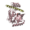
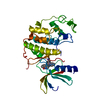
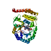
| Title | Crystal Structure of Phenolic Acid Decarboxylase from Bacillus pumilus UI-670 | ||||||
 Components Components | Ferulate decarboxylase | ||||||
 Keywords Keywords | LYASE / beta barrel / lipocalin / biocatalysis / decarboxylase | ||||||
| Function / homology | Phenolic acid decarboxylase / Phenolic acid decarboxylase (PAD) / carboxy-lyase activity / Calycin beta-barrel core domain / Calycin / Lipocalin / Beta Barrel / Mainly Beta / Ferulate decarboxylase Function and homology information Function and homology information | ||||||
| Biological species |  | ||||||
| Method |  X-RAY DIFFRACTION / SYNCHROTRON / MOLECULAR REPLACEMENT / Resolution: 1.69 Å X-RAY DIFFRACTION / SYNCHROTRON / MOLECULAR REPLACEMENT / Resolution: 1.69 Å | ||||||
 Authors Authors | Matte, A. / Grosse, S. / Bergeron, H. / Abokitse, K. / Lau, P.C.K. | ||||||
 Citation Citation | Journal: Acta Crystallogr.,Sect.F / Year: 2010 Title: Structural analysis of Bacillus pumilus phenolic acid decarboxylase, a lipocalin-fold enzyme. Authors: Matte, A. / Grosse, S. / Bergeron, H. / Abokitse, K. / Lau, P.C. #1: Journal: Proteins: Struct.,Funct.,Genet. / Year: 2010Title: p-Coumaric acid decarboxylase from Lactobacillus plantarum: Structural insights into the active site and decarboxylation catalytic mechanism Authors: Rodriguez, H. / Angulo, I. / de las Rivas, B. / Campillo, N. / Paez, J.A. / Munoz, R. / Mancheno, J.M. #2: Journal: Protein Sci. / Year: 1993 Title: Structure and sequence relationships in the lipcalins and related proteins Authors: Flower, D.R. / North, A.C.T. / Attwood, T.K. #4: Journal: J.Bacteriol. / Year: 2008 Title: Phenolic-acid mediated regulation of the padC gene encoding the phenolic acid decarboxylase of Bacillus subtilus Authors: Tran, N.P. / Gury, J. / Dartois, V. / Nguyen, T.K.C. / Seraut, H. / Barthelmebs, L. / Gervais, P. / Cavin, J.-F. #5: Journal: Appl.Environ.Microbiol. / Year: 1995 Title: Cloning, sequencing and expression in Escherichia coli of the Bacillus pumilus gene for ferulic acid decarboxylase Authors: Zago, A. / Degrassi, G. / Bruschi, C.V. | ||||||
| History |
|
- Structure visualization
| Structure viewer | Molecule: MolmilJmol/JSmol |
|---|
- Downloads & links
-Download
| PDBx/mmCIF format | 3nad.cif.gz | 148.8 KB | Display | PDBx/mmCIF format |
|---|---|---|---|---|
| PDB format | pdb3nad.ent.gz | 117.6 KB | Display | PDB format |
| PDBx/mmJSON format | 3nad.json.gz | Tree view | PDBx/mmJSON format | |
| Others |  Other downloads Other downloads |
-Validation report
| Arichive directory | https://data.pdbj.org/pub/pdb/validation_reports/na/3nadftp://data.pdbj.org/pub/pdb/validation_reports/na/3nad | HTTPS FTP |
|---|
-Related structure data
| Related structure data |  2p8gS S: Starting model for refinement |
|---|---|
| Similar structure data |


-Links
 PDBj
PDBj

- Assembly
| Deposited unit | 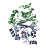
| ||||||||
|---|---|---|---|---|---|---|---|---|---|
| 1 |
| ||||||||
| Unit cell |
|
-Components
| #1: Protein | Mass: 19106.564 Da / Num. of mol.: 2 Source method: isolated from a genetically manipulated source Source: (gene. exp.) #2: Chemical | ChemComp-SO4 / |   Mass: 96.063 Da / Num. of mol.: 1 / Source method: obtained synthetically / Formula: SO4 Mass: 96.063 Da / Num. of mol.: 1 / Source method: obtained synthetically / Formula: SO4#3: Water | ChemComp-HOH / |  Mass: 18.015 Da / Num. of mol.: 368 / Source method: isolated from a natural source / Formula: H2O Mass: 18.015 Da / Num. of mol.: 368 / Source method: isolated from a natural source / Formula: H2O |
|---|
-Experimental details
-Experiment
| Experiment | Method: X-RAY DIFFRACTION / Number of used crystals: 1 |
|---|
- Sample preparation
| Crystal | Density Matthews: 3.01 Å3/Da / Density % sol: 59.14 % |
|---|---|
| Crystal grow | Temperature: 293 K / Method: vapor diffusion / pH: 5.6 Details: 0.2 M Na/K tartarate, 0.1 M tri-sodium citrate, 0.5 M ammonium sulfate, pH 5.6, VAPOR DIFFUSION, temperature 293K |
-Data collection
| Diffraction | Mean temperature: 100 K |
|---|---|
| Diffraction source | Source: SYNCHROTRON / Site: APS  / Beamline: 31-ID / Wavelength: 0.9793 Å / Beamline: 31-ID / Wavelength: 0.9793 Å |
| Detector | Type: Rayonix 225 HE / Detector: CCD / Date: Jun 4, 2008 |
| Radiation | Monochromator: Kohzu HLD-4 double crystal Diamond(111) / Protocol: SINGLE WAVELENGTH / Scattering type: x-ray |
| Radiation wavelength | Wavelength: 0.9793 Å / Relative weight: 1 |
| Reflection | Resolution: 1.69→50 Å / Num. obs: 52226 / % possible obs: 99.1 % / Redundancy: 5.9 % / Rmerge(I) obs: 0.095 / Χ2: 0.976 / Net I/σ(I): 10.5 |
| Reflection shell | Resolution: 1.69→1.75 Å / Redundancy: 4.9 % / Rmerge(I) obs: 0.603 / Num. unique all: 4990 / Χ2: 0.876 / % possible all: 95.7 |
- Processing
| Software |
| |||||||||||||||||||||||||||||||||||||||||||||||||||||||||||||||||||||||||||
|---|---|---|---|---|---|---|---|---|---|---|---|---|---|---|---|---|---|---|---|---|---|---|---|---|---|---|---|---|---|---|---|---|---|---|---|---|---|---|---|---|---|---|---|---|---|---|---|---|---|---|---|---|---|---|---|---|---|---|---|---|---|---|---|---|---|---|---|---|---|---|---|---|---|---|---|---|
| Refinement | Method to determine structure: MOLECULAR REPLACEMENT Starting model: PDB ENTRY 2P8G Resolution: 1.69→50 Å / Cor.coef. Fo:Fc: 0.957 / Cor.coef. Fo:Fc free: 0.949 / WRfactor Rfree: 0.242 / WRfactor Rwork: 0.244 / Occupancy max: 1 / Occupancy min: 0.3 / FOM work R set: 0.876 / SU B: 3.445 / SU ML: 0.056 / SU R Cruickshank DPI: 0.118 / SU Rfree: 0.103 / Cross valid method: THROUGHOUT / σ(F): 0 / ESU R Free: 0.087 / Stereochemistry target values: MAXIMUM LIKELIHOOD / Details: HYDROGENS HAVE BEEN ADDED IN THE RIDING POSITIONS
| |||||||||||||||||||||||||||||||||||||||||||||||||||||||||||||||||||||||||||
| Solvent computation | Ion probe radii: 0.8 Å / Shrinkage radii: 0.8 Å / VDW probe radii: 1.2 Å / Solvent model: MASK | |||||||||||||||||||||||||||||||||||||||||||||||||||||||||||||||||||||||||||
| Displacement parameters | Biso max: 116.32 Å2 / Biso mean: 19.448 Å2 / Biso min: 9.34 Å2
| |||||||||||||||||||||||||||||||||||||||||||||||||||||||||||||||||||||||||||
| Refinement step | Cycle: LAST / Resolution: 1.69→50 Å
| |||||||||||||||||||||||||||||||||||||||||||||||||||||||||||||||||||||||||||
| Refine LS restraints |
| |||||||||||||||||||||||||||||||||||||||||||||||||||||||||||||||||||||||||||
| LS refinement shell | Resolution: 1.69→1.731 Å / Total num. of bins used: 20
| |||||||||||||||||||||||||||||||||||||||||||||||||||||||||||||||||||||||||||
| Refinement TLS params. | Method: refined / Refine-ID: X-RAY DIFFRACTION
| |||||||||||||||||||||||||||||||||||||||||||||||||||||||||||||||||||||||||||
| Refinement TLS group |
|
