Mass: 18.015 Da / Num. of mol.: 1191 / Source method: isolated from a natural source / Formula: H2O
Has protein modification
Y
Sequence details
THE CONSTRUCT (RESIDUES 25-384) WAS EXPRESSED WITH A PURIFICATION TAG MGSDKIHHHHHHENLYFQG. THE TAG ...THE CONSTRUCT (RESIDUES 25-384) WAS EXPRESSED WITH A PURIFICATION TAG MGSDKIHHHHHHENLYFQG. THE TAG WAS REMOVED WITH TEV PROTEASE LEAVING ONLY A GLYCINE (0) FOLLOWED BY THE TARGET SEQUENCE.
-
Experimental details
-
Experiment
Experiment
Method: X-RAY DIFFRACTION / Number of used crystals: 1
-
Sample preparation
Crystal
Density Matthews: 2.34 Å3/Da / Density % sol: 47.33 % Description: DATA WERE SCALED USING XSCALE WITH FRIEDEL PAIRS KEPT AS SEPARATE WHEN COMPUTING R-SYM, COMPLETENESS AND
Crystal grow
Temperature: 277 K / Method: vapor diffusion, sitting drop / pH: 7.7 Details: 0.20M MgAcetate, 20.00% PEG-3350, No Buffer pH 7.7, NANODROP, VAPOR DIFFUSION, SITTING DROP, temperature 277K
Monochromator: SINGLE CRYSTAL SI(111) BENT MONOCHROMATOR (HORIZONTAL FOCUSING) Protocol: MAD / Monochromatic (M) / Laue (L): M / Scattering type: x-ray
Radiation wavelength
ID
Wavelength (Å)
Relative weight
1
0.91837
1
2
0.97947
1
3
0.97901
1
Reflection
Resolution: 1.44→29.738 Å / Num. obs: 132203 / % possible obs: 94.5 % / Observed criterion σ(I): -3 / Biso Wilson estimate: 17.26 Å2 / Rmerge(I) obs: 0.046 / Net I/σ(I): 10.1
Reflection shell
Resolution: 1.44→1.49 Å / Rmerge(I) obs: 0.92 / Mean I/σ(I) obs: 1.2 / % possible all: 88.1
-
Phasing
Phasing
Method: MAD
-
Processing
Software
Name
Version
Classification
NB
MolProbity
3beta29
modelbuilding
PHENIX
refinement
SHELX
phasing
XSCALE
datascaling
PDB_EXTRACT
3.006
dataextraction
XDS
datareduction
SHELXD
phasing
autoSHARP
phasing
BUSTER
2.8.0
refinement
Refinement
Method to determine structure: MAD / Resolution: 1.44→29.738 Å / Cor.coef. Fo:Fc: 0.959 / Cor.coef. Fo:Fc free: 0.953 / Occupancy max: 1 / Occupancy min: 0.2 / Cross valid method: THROUGHOUT / σ(F): 0 Details: 1. A MET-INHIBITION PROTOCOL WAS USED FOR SELENOMETHIONINE INCORPORATION DURING PROTEIN EXPRESSION. THE OCCUPANCY OF THE SE ATOMS IN THE MSE RESIDUES WAS REDUCED TO 0.75 FOR THE REDUCED ...Details: 1. A MET-INHIBITION PROTOCOL WAS USED FOR SELENOMETHIONINE INCORPORATION DURING PROTEIN EXPRESSION. THE OCCUPANCY OF THE SE ATOMS IN THE MSE RESIDUES WAS REDUCED TO 0.75 FOR THE REDUCED SCATTERING POWER DUE TO PARTIAL S-MET INCORPORATION. 2. MAGNESIUM (MG), CHLORIDE (CL), ETHYLENE GLYCOL (EDO) MODELED ARE PRESENT CRYSTALLIZATION/PURIFICATION/CRYO BUFFERS. 3. ATOM RECORD CONTAINS SUM OF TLS AND RESIDUAL B FACTORS. ANISOU RECORD CONTAINS SUM OF TLS AND RESIDUAL U FACTORS. 4. WATERS WERE EXCLUDED FROM AUTOMATIC TLS ASSIGNMENT. 5. RAMACHANDRAN OUTLIER IS LOCATION IN A REGION WITH POOR DENSITY.
Rfactor
Num. reflection
% reflection
Selection details
Rfree
0.19
6661
5.04 %
RANDOM
Rwork
0.167
-
-
-
obs
0.168
132168
-
-
Displacement parameters
Biso mean: 31.56 Å2
Baniso -1
Baniso -2
Baniso -3
1-
4.185 Å2
0 Å2
-4.206 Å2
2-
-
-2.317 Å2
0 Å2
3-
-
-
-1.868 Å2
Refinement step
Cycle: LAST / Resolution: 1.44→29.738 Å
Protein
Nucleic acid
Ligand
Solvent
Total
Num. atoms
5482
0
33
1191
6706
Refine LS restraints
Refine-ID
Type
Dev ideal
Number
Restraint function
Weight
X-RAY DIFFRACTION
t_bond_d
0.01
5921
HARMONIC
2
X-RAY DIFFRACTION
t_angle_deg
0.74
8132
HARMONIC
3.5
X-RAY DIFFRACTION
t_dihedral_angle_d
2067
SINUSOIDAL
2
X-RAY DIFFRACTION
t_incorr_chiral_ct
X-RAY DIFFRACTION
t_pseud_angle
X-RAY DIFFRACTION
t_trig_c_planes
199
HARMONIC
2
X-RAY DIFFRACTION
t_gen_planes
891
HARMONIC
5
X-RAY DIFFRACTION
t_it
5921
HARMONIC
20
X-RAY DIFFRACTION
t_nbd
4
SEMIHARMONIC
5
X-RAY DIFFRACTION
t_omega_torsion
3.25
X-RAY DIFFRACTION
t_other_torsion
15.72
X-RAY DIFFRACTION
t_improper_torsion
X-RAY DIFFRACTION
t_chiral_improper_torsion
796
SEMIHARMONIC
5
X-RAY DIFFRACTION
t_sum_occupancies
X-RAY DIFFRACTION
t_utility_distance
X-RAY DIFFRACTION
t_utility_angle
X-RAY DIFFRACTION
t_utility_torsion
X-RAY DIFFRACTION
t_ideal_dist_contact
8356
SEMIHARMONIC
4
LS refinement shell
Resolution: 1.44→1.48 Å / Total num. of bins used: 20
Rfactor
Num. reflection
% reflection
Rfree
0.27
460
4.91 %
Rwork
0.243
8915
-
all
0.244
9375
-
Refinement TLS params.
Method: refined / Refine-ID: X-RAY DIFFRACTION
ID
L11 (°2)
L12 (°2)
L13 (°2)
L22 (°2)
L23 (°2)
L33 (°2)
S11 (Å °)
S12 (Å °)
S13 (Å °)
S21 (Å °)
S22 (Å °)
S23 (Å °)
S31 (Å °)
S32 (Å °)
S33 (Å °)
T11 (Å2)
T12 (Å2)
T13 (Å2)
T22 (Å2)
T23 (Å2)
T33 (Å2)
Origin x (Å)
Origin y (Å)
Origin z (Å)
1
1.2239
-0.5049
-0.0659
0.5631
0.1948
0.3895
0.0615
0.0117
0.2631
-0.0427
-0.0029
-0.1316
-0.0528
0.0305
-0.0586
-0.0576
0
0.0213
-0.0686
0.0013
0.0161
-4.203
6.922
101.8008
2
3.7366
-1.4581
-2.2654
1.3319
0.692
1.9985
0.1974
0.5194
-0.1841
-0.2853
-0.4056
-0.129
-0.0498
-0.1605
0.2081
-0.173
0.075
0.0835
-0.0635
0.0213
-0.1299
-17.9581
3.0935
70.3671
Refinement TLS group
ID
Refine-ID
Refine TLS-ID
Selection details
Auth asym-ID
Auth seq-ID
1
X-RAY DIFFRACTION
1
{ A|* }
A
33 - 384
2
X-RAY DIFFRACTION
2
{ B|* }
B
31 - 384
+
About Yorodumi
-
News
-
Feb 9, 2022. New format data for meta-information of EMDB entries
New format data for meta-information of EMDB entries
Version 3 of the EMDB header file is now the official format.
The previous official version 1.9 will be removed from the archive.
In the structure databanks used in Yorodumi, some data are registered as the other names, "COVID-19 virus" and "2019-nCoV". Here are the details of the virus and the list of structure data.
Jan 31, 2019. EMDB accession codes are about to change! (news from PDBe EMDB page)
EMDB accession codes are about to change! (news from PDBe EMDB page)
The allocation of 4 digits for EMDB accession codes will soon come to an end. Whilst these codes will remain in use, new EMDB accession codes will include an additional digit and will expand incrementally as the available range of codes is exhausted. The current 4-digit format prefixed with “EMD-” (i.e. EMD-XXXX) will advance to a 5-digit format (i.e. EMD-XXXXX), and so on. It is currently estimated that the 4-digit codes will be depleted around Spring 2019, at which point the 5-digit format will come into force.
The EM Navigator/Yorodumi systems omit the EMD- prefix.
Related info.:Q: What is EMD? / ID/Accession-code notation in Yorodumi/EM Navigator
Yorodumi is a browser for structure data from EMDB, PDB, SASBDB, etc.
This page is also the successor to EM Navigator detail page, and also detail information page/front-end page for Omokage search.
The word "yorodu" (or yorozu) is an old Japanese word meaning "ten thousand". "mi" (miru) is to see.
Related info.:EMDB / PDB / SASBDB / Comparison of 3 databanks / Yorodumi Search / Aug 31, 2016. New EM Navigator & Yorodumi / Yorodumi Papers / Jmol/JSmol / Function and homology information / Changes in new EM Navigator and Yorodumi
 Movie
Movie Controller

 Yorodumi
 Open data
 Basic information
 Components
 Keywords
 Function and homology information
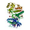
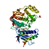
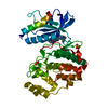
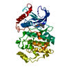
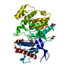
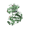
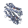
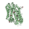
 Bacteroides ovatus (bacteria)
 X-RAY DIFFRACTION /
  Authors
 Citation
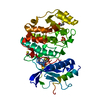
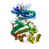
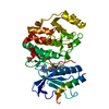
 Structure visualization
 Downloads & links
 Other downloads
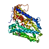

 PDBj
PDBj


 Assembly
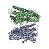

 Mass: 24.305 Da / Num. of mol.: 3 / Source method: obtained synthetically / Formula: Mg
 Mass: 35.453 Da / Num. of mol.: 2 / Source method: obtained synthetically / Formula: Cl
 Mass: 62.068 Da / Num. of mol.: 7 / Source method: obtained synthetically / Formula: C2H6O2
 Mass: 18.015 Da / Num. of mol.: 1191 / Source method: isolated from a natural source / Formula: H2O
 Sample preparation
 / Beamline: BL11-1 / Wavelength: 0.91837,0.97947,0.97901
 Processing