[日本語] English
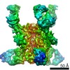
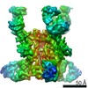
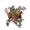
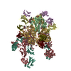
- PDB-3n6r: CRYSTAL STRUCTURE OF the holoenzyme of PROPIONYL-COA CARBOXYLASE (PCC) -

+
データを開く


IDまたはキーワード:

読み込み中...

-
基本情報

登録情報
データベース: PDB / ID: 3n6r
タイトルCRYSTAL STRUCTURE OF the holoenzyme of PROPIONYL-COA CARBOXYLASE (PCC)
要素
  • Propionyl-CoA carboxylase, alpha subunit
  • Propionyl-CoA carboxylase, beta subunit
キーワードLIGASE / protein complex / biotin-dependent carboxylase
機能・相同性
機能・相同性情報


propionate metabolic process / propionyl-CoA carboxylase / propionyl-CoA carboxylase activity / lipid catabolic process / ATP binding / metal ion binding
類似検索 - 分子機能
Glycoprotein, Type 4 Pilin - #30 / Propionyl-coenzyme A carboxylase, BT domain / Propionyl-coenzyme A carboxylase BT domain / Glycoprotein, Type 4 Pilin / Acetyl-coenzyme A carboxyltransferase, C-terminal / Acetyl-coenzyme A (CoA) carboxyltransferase C-terminal domain profile. / Acetyl-coenzyme A carboxyltransferase, N-terminal / Acetyl-coenzyme A (CoA) carboxyltransferase N-terminal domain profile. / Acetyl-CoA carboxylase / Carboxyl transferase domain ...Glycoprotein, Type 4 Pilin - #30 / Propionyl-coenzyme A carboxylase, BT domain / Propionyl-coenzyme A carboxylase BT domain / Glycoprotein, Type 4 Pilin / Acetyl-coenzyme A carboxyltransferase, C-terminal / Acetyl-coenzyme A (CoA) carboxyltransferase C-terminal domain profile. / Acetyl-coenzyme A carboxyltransferase, N-terminal / Acetyl-coenzyme A (CoA) carboxyltransferase N-terminal domain profile. / Acetyl-CoA carboxylase / Carboxyl transferase domain / Biotin-binding site / Biotin-requiring enzymes attachment site. / Biotin carboxylase-like, N-terminal domain / Biotin carboxylase, C-terminal / Biotin carboxylation domain / Biotin carboxylase, N-terminal domain / Biotin carboxylase C-terminal domain / Biotin carboxylation domain profile. / Biotin carboxylase C-terminal domain / Rossmann fold - #20 / Carbamoyl-phosphate synthase subdomain signature 1. / RNA polymerase II/Efflux pump adaptor protein, barrel-sandwich hybrid domain / Carbamoyl-phosphate synthetase large subunit-like, ATP-binding domain / Carbamoyl-phosphate synthase L chain, ATP binding domain / Biotin-requiring enzyme / ATP-grasp fold, A domain / Rudiment single hybrid motif / Biotinyl/lipoyl domain profile. / Biotin/lipoyl attachment / Single hybrid motif / 2-enoyl-CoA Hydratase; Chain A, domain 1 / 2-enoyl-CoA Hydratase; Chain A, domain 1 / ATP-grasp fold, B domain / ATP-grasp fold, subdomain 1 / Pre-ATP-grasp domain superfamily / ATP-grasp fold / ATP-grasp fold profile. / D-amino Acid Aminotransferase; Chain A, domain 1 / ClpP/crotonase-like domain superfamily / Dna Ligase; domain 1 / OB fold (Dihydrolipoamide Acetyltransferase, E2P) / Carbamoyl-phosphate synthase subdomain signature 2. / Alpha-Beta Complex / Beta Barrel / Rossmann fold / 2-Layer Sandwich / 3-Layer(aba) Sandwich / Mainly Beta / Alpha Beta
類似検索 - ドメイン・相同性
Chem-BTI / Propionyl-CoA carboxylase beta chain / Propionyl-CoA carboxylase alpha chain
類似検索 - 構成要素
生物種Ruegeria pomeroyi (バクテリア)
Roseobacter denitrificans (バクテリア)
手法X線回折 / シンクロトロン / 分子置換 / 解像度: 3.2 Å
データ登録者Huang, C.S. / Sadre-Bazzaz, K. / Tong, L.
引用ジャーナル: Nature / : 2010
タイトル: Crystal structure of the alpha(6)beta(6) holoenzyme of propionyl-coenzyme A carboxylase.
著者: Christine S Huang / Kianoush Sadre-Bazzaz / Yang Shen / Binbin Deng / Z Hong Zhou / Liang Tong /
要旨: Propionyl-coenzyme A carboxylase (PCC), a mitochondrial biotin-dependent enzyme, is essential for the catabolism of the amino acids Thr, Val, Ile and Met, cholesterol and fatty acids with an odd ...Propionyl-coenzyme A carboxylase (PCC), a mitochondrial biotin-dependent enzyme, is essential for the catabolism of the amino acids Thr, Val, Ile and Met, cholesterol and fatty acids with an odd number of carbon atoms. Deficiencies in PCC activity in humans are linked to the disease propionic acidaemia, an autosomal recessive disorder that can be fatal in infants. The holoenzyme of PCC is an alpha(6)beta(6) dodecamer, with a molecular mass of 750 kDa. The alpha-subunit contains the biotin carboxylase (BC) and biotin carboxyl carrier protein (BCCP) domains, whereas the beta-subunit supplies the carboxyltransferase (CT) activity. Here we report the crystal structure at 3.2-A resolution of a bacterial PCC alpha(6)beta(6) holoenzyme as well as cryo-electron microscopy (cryo-EM) reconstruction at 15-A resolution demonstrating a similar structure for human PCC. The structure defines the overall architecture of PCC and reveals unexpectedly that the alpha-subunits are arranged as monomers in the holoenzyme, decorating a central beta(6) hexamer. A hitherto unrecognized domain in the alpha-subunit, formed by residues between the BC and BCCP domains, is crucial for interactions with the beta-subunit. We have named it the BT domain. The structure reveals for the first time the relative positions of the BC and CT active sites in the holoenzyme. They are separated by approximately 55 A, indicating that the entire BCCP domain must translocate during catalysis. The BCCP domain is located in the active site of the beta-subunit in the current structure, providing insight for its involvement in the CT reaction. The structural information establishes a molecular basis for understanding the large collection of disease-causing mutations in PCC and is relevant for the holoenzymes of other biotin-dependent carboxylases, including 3-methylcrotonyl-CoA carboxylase (MCC) and eukaryotic acetyl-CoA carboxylase (ACC).
履歴
登録2010年5月26日登録サイト: RCSB / 処理サイト: RCSB
改定 1.02010年8月25日Provider: repository / タイプ: Initial release
改定 1.12011年7月13日Group: Source and taxonomy / Version format compliance

-
構造の表示

構造ビューア分子:
MolmilJmol/JSmol

ダウンロードとリンク

-
集合体

登録構造単位
A: Propionyl-CoA carboxylase, alpha subunit
B: Propionyl-CoA carboxylase, beta subunit
C: Propionyl-CoA carboxylase, alpha subunit
D: Propionyl-CoA carboxylase, beta subunit
E: Propionyl-CoA carboxylase, alpha subunit
F: Propionyl-CoA carboxylase, beta subunit
G: Propionyl-CoA carboxylase, alpha subunit
H: Propionyl-CoA carboxylase, beta subunit
I: Propionyl-CoA carboxylase, alpha subunit
J: Propionyl-CoA carboxylase, beta subunit
K: Propionyl-CoA carboxylase, alpha subunit
L: Propionyl-CoA carboxylase, beta subunit
ヘテロ分子


分子量 (理論値)分子数
合計 (水以外)795,86218
ポリマ-794,49212
非ポリマー1,3706
00
1


  • 登録構造と同一
  • 登録者・ソフトウェアが定義した集合体
タイプ名称対称操作
identity operation1_555x,y,z1
Buried area73220 Å2
ΔGint-286 kcal/mol
Surface area237170 Å2
手法PISA
単位格子
Length a, b, c (Å)133.890, 159.170, 153.740
Angle α, β, γ (deg.)113.87, 101.03, 108.99
Int Tables number1
Space group name H-MP1

-
要素

#1: タンパク質
Propionyl-CoA carboxylase, alpha subunit


分子量: 73946.180 Da / 分子数: 6 / 由来タイプ: 組換発現 / 由来: (組換発現) Ruegeria pomeroyi (バクテリア) / 遺伝子: pccA, SPO1101 / 発現宿主: Escherichia coli (大腸菌) / 参照: UniProt: Q5LUF3, propionyl-CoA carboxylase
#2: タンパク質
Propionyl-CoA carboxylase, beta subunit


分子量: 58469.172 Da / 分子数: 6 / 由来タイプ: 組換発現
由来: (組換発現) Roseobacter denitrificans (バクテリア)
: OCh 114 / 遺伝子: pccB, RD1_2028 / 発現宿主: Escherichia coli (大腸菌) / 参照: UniProt: Q168G2, propionyl-CoA carboxylase
#3: 化合物
ChemComp-BTI / 5-(HEXAHYDRO-2-OXO-1H-THIENO[3,4-D]IMIDAZOL-6-YL)PENTANAL


分子量: 228.311 Da / 分子数: 6 / 由来タイプ: 合成 / : C10H16N2O2S

-
実験情報

-
実験

実験手法: X線回折 / 使用した結晶の数: 1

-
試料調製

結晶マシュー密度: 3.37 Å3/Da / 溶媒含有率: 63.53 %

-
データ収集

回折平均測定温度: 100 K
放射光源由来: シンクロトロン / サイト: NSLS / ビームライン: X29A / 波長: 1.0809 Å
検出器タイプ: ADSC QUANTUM 315 / 検出器: CCD / 日付: 2009年7月31日 / 詳細: MIRRORS
放射モノクロメーター: SI(111) / プロトコル: SINGLE WAVELENGTH / 単色(M)・ラウエ(L): M / 散乱光タイプ: x-ray
放射波長波長: 1.0809 Å / 相対比: 1
反射解像度: 3.2→30 Å / Num. obs: 169648 / % possible obs: 92 % / 冗長度: 1.9 % / Rmerge(I) obs: 0.084 / Net I/σ(I): 8.6854
反射 シェル解像度: 3.2→3.31 Å / 冗長度: 1.7 % / Rmerge(I) obs: 0.342 / Mean I/σ(I) obs: 2.024 / % possible all: 80

-
解析

ソフトウェア
名称バージョン分類
CBASSデータ収集
PHASER位相決定
CNS1.2精密化
DENZOデータ削減
SCALEPACKデータスケーリング
精密化構造決定の手法: 分子置換 / 解像度: 3.2→29.36 Å / Rfactor Rfree error: 0.002 / Data cutoff high absF: 194743.4 / Data cutoff low absF: 0 / Isotropic thermal model: RESTRAINED / 交差検証法: THROUGHOUT / σ(F): 0
詳細: BULK SOLVENT MODEL USED. THE RESIDUES ARE NUMBERED ACCORDING TO THE HUMAN PCC SEQUENCE
Rfactor反射数%反射Selection details
Rfree0.245 11579 7.5 %RANDOM
Rwork0.212 ---
obs0.212 155003 92.4 %-
溶媒の処理溶媒モデル: FLAT MODEL / Bsol: 26.3531 Å2 / ksol: 0.3 e/Å3
原子変位パラメータBiso mean: 51.2 Å2
Baniso -1Baniso -2Baniso -3
1-3.9 Å21.03 Å2-4.3 Å2
2---1.42 Å2-8.64 Å2
3----2.48 Å2
Refine analyze
FreeObs
Luzzati coordinate error0.41 Å0.35 Å
Luzzati d res low-5 Å
Luzzati sigma a0.63 Å0.57 Å
精密化ステップサイクル: LAST / 解像度: 3.2→29.36 Å
タンパク質核酸リガンド溶媒全体
原子数51831 0 90 0 51921
拘束条件
Refine-IDタイプDev ideal
X-RAY DIFFRACTIONc_bond_d0.012
X-RAY DIFFRACTIONc_bond_d_na
X-RAY DIFFRACTIONc_bond_d_prot
X-RAY DIFFRACTIONc_angle_d
X-RAY DIFFRACTIONc_angle_d_na
X-RAY DIFFRACTIONc_angle_d_prot
X-RAY DIFFRACTIONc_angle_deg1.6
X-RAY DIFFRACTIONc_angle_deg_na
X-RAY DIFFRACTIONc_angle_deg_prot
X-RAY DIFFRACTIONc_dihedral_angle_d23.9
X-RAY DIFFRACTIONc_dihedral_angle_d_na
X-RAY DIFFRACTIONc_dihedral_angle_d_prot
X-RAY DIFFRACTIONc_improper_angle_d1.07
X-RAY DIFFRACTIONc_improper_angle_d_na
X-RAY DIFFRACTIONc_improper_angle_d_prot
X-RAY DIFFRACTIONc_mcbond_it
X-RAY DIFFRACTIONc_mcangle_it
X-RAY DIFFRACTIONc_scbond_it
X-RAY DIFFRACTIONc_scangle_it
Refine LS restraints NCSNCS model details: NONE
LS精密化 シェル解像度: 3.2→3.31 Å / Rfactor Rfree error: 0.011 / Total num. of bins used: 10
Rfactor反射数%反射
Rfree0.34 1038 7.7 %
Rwork0.319 12393 -
obs--80 %
Xplor file
Refine-IDSerial noParam fileTopol file
X-RAY DIFFRACTION1protein_rep.paramprotein.top
X-RAY DIFFRACTION2bti.par

+
万見について

-
お知らせ

-
2022年2月9日: EMDBエントリの付随情報ファイルのフォーマットが新しくなりました

EMDBエントリの付随情報ファイルのフォーマットが新しくなりました

  • EMDBのヘッダファイルのバージョン3が、公式のフォーマットとなりました。
  • これまでは公式だったバージョン1.9は、アーカイブから削除されます。

関連情報:EMDBヘッダ

外部リンク:wwPDBはEMDBデータモデルのバージョン3へ移行します

-
2020年8月12日: 新型コロナ情報

新型コロナ情報

URL: https://pdbjlvh1.pdbj.org/emnavi/covid19.php

新ページ: EM Navigatorに新型コロナウイルスの特設ページを開設しました。

関連情報:Covid-19情報 / 2020年3月5日: 新型コロナウイルスの構造データ

+
2020年3月5日: 新型コロナウイルスの構造データ

新型コロナウイルスの構造データ

関連情報:万見生物種 / 2020年8月12日: 新型コロナ情報

外部リンク:COVID-19特集ページ - PDBj / 今月の分子2020年2月:コロナウイルスプロテーアーゼ

+
2019年1月31日: EMDBのIDの桁数の変更

EMDBのIDの桁数の変更

  • EMDBエントリに付与されているアクセスコード(EMDB-ID)は4桁の数字(例、EMD-1234)でしたが、間もなく枯渇します。これまでの4桁のID番号は4桁のまま変更されませんが、4桁の数字を使い切った後に発行されるIDは5桁以上の数字(例、EMD-12345)になります。5桁のIDは2019年の春頃から発行される見通しです。
  • EM Navigator/万見では、接頭語「EMD-」は省略されています。

関連情報:Q: 「EMD」とは何ですか? / 万見/EM NavigatorにおけるID/アクセスコードの表記

外部リンク:EMDB Accession Codes are Changing Soon! / PDBjへお問い合わせ

+
2017年7月12日: PDB大規模アップデート

PDB大規模アップデート

  • 新バージョンのPDBx/mmCIF辞書形式に基づくデータがリリースされました。
  • 今回の更新はバージョン番号が4から5になる大規模なもので、全エントリデータの書き換えが行われる「Remediation」というアップデートに該当します。
  • このバージョンアップで、電子顕微鏡の実験手法に関する多くの項目の書式が改定されました(例:em_softwareなど)。
  • EM NavigatorとYorodumiでも、この改定に基づいた表示内容になります。

外部リンク:wwPDB Remediation / OneDepデータ基準に準拠した、より強化された内容のモデル構造ファイルが、PDBアーカイブで公開されました。

-
万見 (Yorodumi)

幾万の構造データを、幾万の視点から

  • 万見(Yorodumi)は、EMDB/PDB/SASBDBなどの構造データを閲覧するためのページです。
  • EM Navigatorの詳細ページの後継、Omokage検索のフロントエンドも兼ねています。

関連情報:EMDB / PDB / SASBDB / 3つのデータバンクの比較 / 万見検索 / 2016年8月31日: 新しいEM Navigatorと万見 / 万見文献 / Jmol/JSmol / 機能・相同性情報 / 新しいEM Navigatorと万見の変更点

他の情報も見る