 Movie
Movie Controller


+ Open data

- Basic information
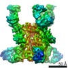
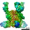
| Entry | Database: EMDB / ID: EMD-5345 | |||||||||
|---|---|---|---|---|---|---|---|---|---|---|
| Title | CryoEM Structure of the Human Propionyl-CoA Carboxylase | |||||||||
 Map data Map data | This is the volume. | |||||||||
 Sample Sample |
| |||||||||
 Keywords Keywords | CryoEM / human propionyl-CoA carboxylase / three-dimensional structure / single particle analysis | |||||||||
| Biological species |  Homo sapiens (human) Homo sapiens (human) | |||||||||
| Method | single particle reconstruction / cryo EM / Resolution: 15.0 Å | |||||||||
 Authors Authors | Zhou ZH / Deng B / Shen Y / Tong L | |||||||||
 Citation Citation | Journal: Nature / Year: 2010 Title: Crystal structure of the alpha(6)beta(6) holoenzyme of propionyl-coenzyme A carboxylase. Authors: Christine S Huang / Kianoush Sadre-Bazzaz / Yang Shen / Binbin Deng / Z Hong Zhou / Liang Tong /  Abstract: Propionyl-coenzyme A carboxylase (PCC), a mitochondrial biotin-dependent enzyme, is essential for the catabolism of the amino acids Thr, Val, Ile and Met, cholesterol and fatty acids with an odd ...Propionyl-coenzyme A carboxylase (PCC), a mitochondrial biotin-dependent enzyme, is essential for the catabolism of the amino acids Thr, Val, Ile and Met, cholesterol and fatty acids with an odd number of carbon atoms. Deficiencies in PCC activity in humans are linked to the disease propionic acidaemia, an autosomal recessive disorder that can be fatal in infants. The holoenzyme of PCC is an alpha(6)beta(6) dodecamer, with a molecular mass of 750 kDa. The alpha-subunit contains the biotin carboxylase (BC) and biotin carboxyl carrier protein (BCCP) domains, whereas the beta-subunit supplies the carboxyltransferase (CT) activity. Here we report the crystal structure at 3.2-A resolution of a bacterial PCC alpha(6)beta(6) holoenzyme as well as cryo-electron microscopy (cryo-EM) reconstruction at 15-A resolution demonstrating a similar structure for human PCC. The structure defines the overall architecture of PCC and reveals unexpectedly that the alpha-subunits are arranged as monomers in the holoenzyme, decorating a central beta(6) hexamer. A hitherto unrecognized domain in the alpha-subunit, formed by residues between the BC and BCCP domains, is crucial for interactions with the beta-subunit. We have named it the BT domain. The structure reveals for the first time the relative positions of the BC and CT active sites in the holoenzyme. They are separated by approximately 55 A, indicating that the entire BCCP domain must translocate during catalysis. The BCCP domain is located in the active site of the beta-subunit in the current structure, providing insight for its involvement in the CT reaction. The structural information establishes a molecular basis for understanding the large collection of disease-causing mutations in PCC and is relevant for the holoenzymes of other biotin-dependent carboxylases, including 3-methylcrotonyl-CoA carboxylase (MCC) and eukaryotic acetyl-CoA carboxylase (ACC). | |||||||||
| History |
|
- Structure visualization
| Movie |
 Movie viewer Movie viewer |
|---|---|
| Structure viewer | EM map: SurfViewMolmilJmol/JSmol |
| Supplemental images |
 UCSF Chimera
- Downloads & links
-EMDB archive
| Map data | emd_5345.map.gz | 4.3 MB | EMDB map data format | |
|---|---|---|---|---|
| Header (meta data) | emd-5345-v30.xmlemd-5345.xml | 10.2 KB 10.2 KB | Display Display | EMDB header |
| Images |  emd_5345_1.png emd_5345_1.png | 111.5 KB | ||
| Archive directory |  http://ftp.pdbj.org/pub/emdb/structures/EMD-5345ftp://ftp.pdbj.org/pub/emdb/structures/EMD-5345 http://ftp.pdbj.org/pub/emdb/structures/EMD-5345ftp://ftp.pdbj.org/pub/emdb/structures/EMD-5345 | HTTPS FTP |
-Related structure data


-Links
| EMDB pages | EMDB (EBI/PDBe) / EMDataResource |
|---|
-Map
| File | Download / File: emd_5345.map.gz / Format: CCP4 / Size: 5.2 MB / Type: IMAGE STORED AS FLOATING POINT NUMBER (4 BYTES) | ||||||||||||||||||||||||||||||||||||||||||||||||||||||||||||||||||||
|---|---|---|---|---|---|---|---|---|---|---|---|---|---|---|---|---|---|---|---|---|---|---|---|---|---|---|---|---|---|---|---|---|---|---|---|---|---|---|---|---|---|---|---|---|---|---|---|---|---|---|---|---|---|---|---|---|---|---|---|---|---|---|---|---|---|---|---|---|---|
| Annotation | This is the volume. | ||||||||||||||||||||||||||||||||||||||||||||||||||||||||||||||||||||
| Projections & slices | Image control
Images are generated by Spider. | ||||||||||||||||||||||||||||||||||||||||||||||||||||||||||||||||||||
| Voxel size | X=Y=Z: 2.8 Å | ||||||||||||||||||||||||||||||||||||||||||||||||||||||||||||||||||||
| Density |
| ||||||||||||||||||||||||||||||||||||||||||||||||||||||||||||||||||||
| Symmetry | Space group: 1 | ||||||||||||||||||||||||||||||||||||||||||||||||||||||||||||||||||||
| Details | EMDB XML:
CCP4 map header:
| ||||||||||||||||||||||||||||||||||||||||||||||||||||||||||||||||||||
 Z (Sec.)
 Y (Row.)
 X (Col.)


-Supplemental data
- Sample components
-Entire : Human Propionyl-CoA Carboxylase holoenzyme
| Entire | Name: Human Propionyl-CoA Carboxylase holoenzyme |
|---|---|
| Components |
|
-Supramolecule #1000: Human Propionyl-CoA Carboxylase holoenzyme
| Supramolecule | Name: Human Propionyl-CoA Carboxylase holoenzyme / type: sample / ID: 1000 / Details: The sample was monodisperse Oligomeric state: dodecamer (six alpha and six beta subunits) Number unique components: 2 |
|---|---|
| Molecular weight | Experimental: 750 KDa / Theoretical: 768 KDa / Method: SDS PAGE |
-Macromolecule #1: Human Propionyl-CoA Carboxylase
| Macromolecule | Name: Human Propionyl-CoA Carboxylase / type: protein_or_peptide / ID: 1 / Name.synonym: Human Propionyl-CoA Carboxylase / Details: PCC in solution / Number of copies: 6 / Oligomeric state: dodecamer / Recombinant expression: Yes |
|---|---|
| Source (natural) | Organism: Homo sapiens (human) / synonym: Human / Location in cell: cytoplasm |
| Molecular weight | Experimental: 750 KDa / Theoretical: 768 KDa |
| Recombinant expression | Organism:  |
-Experimental details
-Structure determination
| Method | cryo EM |
|---|---|
 Processing Processing | single particle reconstruction |
| Aggregation state | particle |
-Sample preparation
| Concentration | 0.07 mg/mL |
|---|---|
| Buffer | pH: 7.4 / Details: 25 mM Tris, 250 mM NaCl, 2 mM DTT, 5% v/v glycerol |
| Grid | Details: 300 mesh copper quantifoil grid |
| Vitrification | Cryogen name: NITROGEN / Chamber humidity: 40 % / Chamber temperature: 90 K / Instrument: HOMEMADE PLUNGER / Details: Vitrification instrument: home-made plunger / Method: blot for 2 seconds before plunging |
- Electron microscopy
| Microscope | JEOL 1200EX |
|---|---|
| Temperature | Min: 77 K / Max: 100 K / Average: 90 K |
| Alignment procedure | Legacy - Astigmatism: corrected at 100,000 times mag |
| Image recording | Category: FILM / Film or detector model: KODAK SO-163 FILM / Digitization - Scanner: ZEISS SCAI / Digitization - Sampling interval: 14 µm / Average electron dose: 10 e/Å2 / Bits/pixel: 12 |
| Electron beam | Acceleration voltage: 100 kV / Electron source: LAB6 |
| Electron optics | Calibrated magnification: 50000 / Illumination mode: FLOOD BEAM / Imaging mode: BRIGHT FIELD / Cs: 4.1 mm / Nominal defocus max: 1.5 µm / Nominal defocus min: 0.5 µm / Nominal magnification: 50000 |
| Sample stage | Specimen holder: eucentric / Specimen holder model: GATAN LIQUID NITROGEN |
-Image processing
| CTF correction | Details: each particle |
|---|---|
| Final reconstruction | Algorithm: OTHER / Resolution.type: BY AUTHOR / Resolution: 15.0 Å / Resolution method: FSC 0.5 CUT-OFF / Software - Name: EMAN / Number images used: 1000 |
